Zinc »
PDB 5gm3-5h7r »
5h4g »
Zinc in PDB 5h4g: Structure of Pin-Domain Protein (VAPC4 Toxin) From Pyrococcus Horikoshii Determined at 1.77 A Resolution
Protein crystallography data
The structure of Structure of Pin-Domain Protein (VAPC4 Toxin) From Pyrococcus Horikoshii Determined at 1.77 A Resolution, PDB code: 5h4g
was solved by
A.Biswas,
K.Hatti,
N.Srinivasan,
M.R.N.Murthy,
K.Sekar,
with X-Ray Crystallography technique. A brief refinement statistics is given in the table below:
| Resolution Low / High (Å) | 50.94 / 1.77 |
| Space group | P 1 21 1 |
| Cell size a, b, c (Å), α, β, γ (°) | 50.780, 44.640, 52.370, 90.00, 103.40, 90.00 |
| R / Rfree (%) | 16.7 / 22.1 |
Zinc Binding Sites:
The binding sites of Zinc atom in the Structure of Pin-Domain Protein (VAPC4 Toxin) From Pyrococcus Horikoshii Determined at 1.77 A Resolution
(pdb code 5h4g). This binding sites where shown within
5.0 Angstroms radius around Zinc atom.
In total only one binding site of Zinc was determined in the Structure of Pin-Domain Protein (VAPC4 Toxin) From Pyrococcus Horikoshii Determined at 1.77 A Resolution, PDB code: 5h4g:
In total only one binding site of Zinc was determined in the Structure of Pin-Domain Protein (VAPC4 Toxin) From Pyrococcus Horikoshii Determined at 1.77 A Resolution, PDB code: 5h4g:
Zinc binding site 1 out of 1 in 5h4g
Go back to
Zinc binding site 1 out
of 1 in the Structure of Pin-Domain Protein (VAPC4 Toxin) From Pyrococcus Horikoshii Determined at 1.77 A Resolution
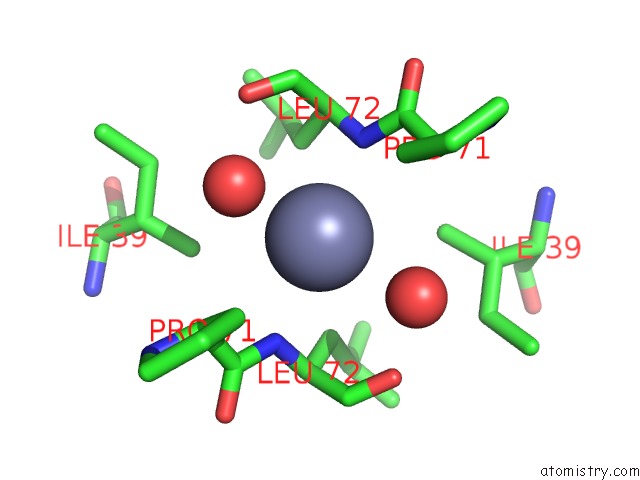
Mono view
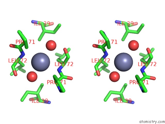
Stereo pair view
Mono view
Stereo pair view
A full contact list of Zinc with other atoms in the Zn binding
site number 1 of Structure of Pin-Domain Protein (VAPC4 Toxin) From Pyrococcus Horikoshii Determined at 1.77 A Resolution within 5.0Å range:
|
Reference:
K.Hatti,
A.Biswas,
S.Chaudhary,
V.Dadireddy,
K.Sekar,
N.Srinivasan,
M.R.N.Murthy.
Structure Determination of Contaminant Proteins Using the Marathonmr Procedure J. Struct. Biol. V. 197 372 2017.
ISSN: ESSN 1095-8657
PubMed: 28167161
DOI: 10.1016/J.JSB.2017.01.005
Page generated: Sun Oct 27 17:19:21 2024
ISSN: ESSN 1095-8657
PubMed: 28167161
DOI: 10.1016/J.JSB.2017.01.005
Last articles
Zn in 9MJ5Zn in 9HNW
Zn in 9G0L
Zn in 9FNE
Zn in 9DZN
Zn in 9E0I
Zn in 9D32
Zn in 9DAK
Zn in 8ZXC
Zn in 8ZUF