Zinc »
PDB 5gm3-5h7r »
5gwd »
Zinc in PDB 5gwd: Structure of Myroilysin
Protein crystallography data
The structure of Structure of Myroilysin, PDB code: 5gwd
was solved by
D.Xu,
T.Ran,
W.Wang,
with X-Ray Crystallography technique. A brief refinement statistics is given in the table below:
| Resolution Low / High (Å) | 19.90 / 1.89 |
| Space group | P 1 21 1 |
| Cell size a, b, c (Å), α, β, γ (°) | 72.223, 35.486, 93.757, 90.00, 101.41, 90.00 |
| R / Rfree (%) | 18.2 / 22.4 |
Zinc Binding Sites:
The binding sites of Zinc atom in the Structure of Myroilysin
(pdb code 5gwd). This binding sites where shown within
5.0 Angstroms radius around Zinc atom.
In total 2 binding sites of Zinc where determined in the Structure of Myroilysin, PDB code: 5gwd:
Jump to Zinc binding site number: 1; 2;
In total 2 binding sites of Zinc where determined in the Structure of Myroilysin, PDB code: 5gwd:
Jump to Zinc binding site number: 1; 2;
Zinc binding site 1 out of 2 in 5gwd
Go back to
Zinc binding site 1 out
of 2 in the Structure of Myroilysin
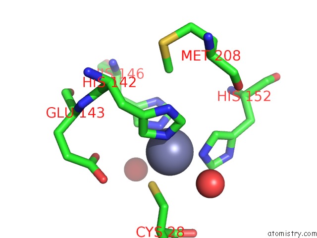
Mono view
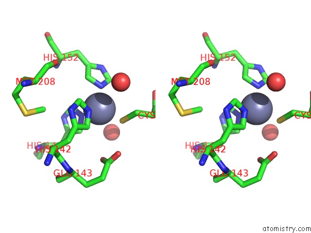
Stereo pair view
Mono view
Stereo pair view
A full contact list of Zinc with other atoms in the Zn binding
site number 1 of Structure of Myroilysin within 5.0Å range:
|
Zinc binding site 2 out of 2 in 5gwd
Go back to
Zinc binding site 2 out
of 2 in the Structure of Myroilysin
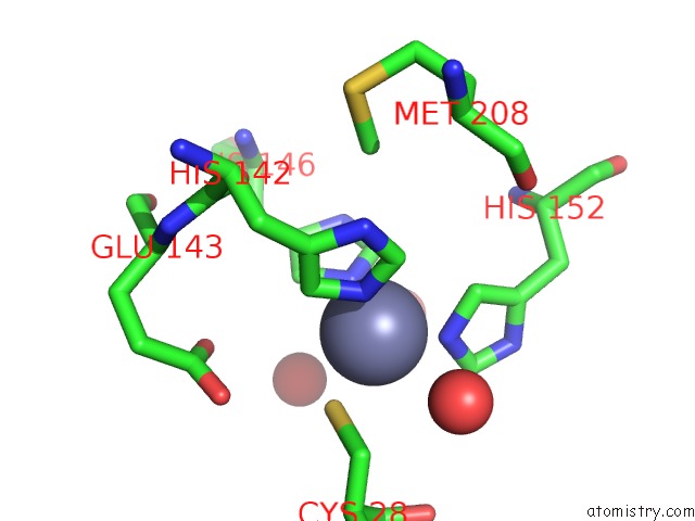
Mono view
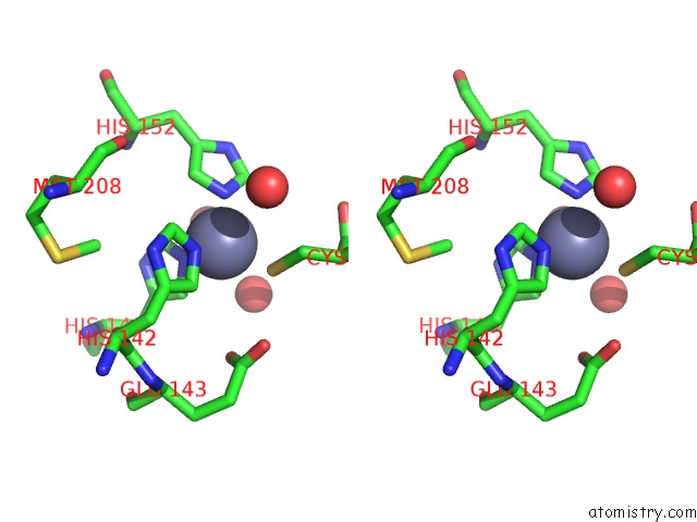
Stereo pair view
Mono view
Stereo pair view
A full contact list of Zinc with other atoms in the Zn binding
site number 2 of Structure of Myroilysin within 5.0Å range:
|
Reference:
D.Xu,
J.Zhou,
X.Lou,
J.He,
T.Ran,
W.Wang.
Myroilysin Is A New Bacterial Member of the M12A Family of Metzincin Metallopeptidases and Is Activated By A Cysteine Switch Mechanism. J. Biol. Chem. V. 292 5195 2017.
ISSN: ESSN 1083-351X
PubMed: 28188295
DOI: 10.1074/JBC.M116.758110
Page generated: Sun Oct 27 17:13:55 2024
ISSN: ESSN 1083-351X
PubMed: 28188295
DOI: 10.1074/JBC.M116.758110
Last articles
Zn in 9MJ5Zn in 9HNW
Zn in 9G0L
Zn in 9FNE
Zn in 9DZN
Zn in 9E0I
Zn in 9D32
Zn in 9DAK
Zn in 8ZXC
Zn in 8ZUF