Zinc »
PDB 4yn2-4z1d »
4ywp »
Zinc in PDB 4ywp: Sucrose Binding Site in Genetically Engineered Carbonic Anhydrase IX
Enzymatic activity of Sucrose Binding Site in Genetically Engineered Carbonic Anhydrase IX
All present enzymatic activity of Sucrose Binding Site in Genetically Engineered Carbonic Anhydrase IX:
4.2.1.1;
4.2.1.1;
Protein crystallography data
The structure of Sucrose Binding Site in Genetically Engineered Carbonic Anhydrase IX, PDB code: 4ywp
was solved by
M.A.Pinard,
M.Aggarwal,
with X-Ray Crystallography technique. A brief refinement statistics is given in the table below:
| Resolution Low / High (Å) | 20.00 / 1.45 |
| Space group | P 1 21 1 |
| Cell size a, b, c (Å), α, β, γ (°) | 41.845, 41.240, 72.275, 90.00, 103.76, 90.00 |
| R / Rfree (%) | 16.2 / 19 |
Zinc Binding Sites:
The binding sites of Zinc atom in the Sucrose Binding Site in Genetically Engineered Carbonic Anhydrase IX
(pdb code 4ywp). This binding sites where shown within
5.0 Angstroms radius around Zinc atom.
In total only one binding site of Zinc was determined in the Sucrose Binding Site in Genetically Engineered Carbonic Anhydrase IX, PDB code: 4ywp:
In total only one binding site of Zinc was determined in the Sucrose Binding Site in Genetically Engineered Carbonic Anhydrase IX, PDB code: 4ywp:
Zinc binding site 1 out of 1 in 4ywp
Go back to
Zinc binding site 1 out
of 1 in the Sucrose Binding Site in Genetically Engineered Carbonic Anhydrase IX

Mono view
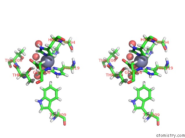
Stereo pair view
Mono view
Stereo pair view
A full contact list of Zinc with other atoms in the Zn binding
site number 1 of Sucrose Binding Site in Genetically Engineered Carbonic Anhydrase IX within 5.0Å range:
|
Reference:
M.A.Pinard,
M.Aggarwal,
B.P.Mahon,
C.Tu,
R.Mckenna.
A Sucrose-Binding Site Provides A Lead Towards An Isoform-Specific Inhibitor of the Cancer-Associated Enzyme Carbonic Anhydrase IX. Acta Crystallogr F Struct V. 71 1352 2015BIOL Commun.
ISSN: ESSN 2053-230X
PubMed: 26457530
DOI: 10.1107/S2053230X1501239X
Page generated: Sun Oct 27 11:28:39 2024
ISSN: ESSN 2053-230X
PubMed: 26457530
DOI: 10.1107/S2053230X1501239X
Last articles
Zn in 9MJ5Zn in 9HNW
Zn in 9G0L
Zn in 9FNE
Zn in 9DZN
Zn in 9E0I
Zn in 9D32
Zn in 9DAK
Zn in 8ZXC
Zn in 8ZUF