Zinc »
PDB 4kjv-4kxq »
4kvp »
Zinc in PDB 4kvp: Human P53 Core Domain Mutant V157F
Protein crystallography data
The structure of Human P53 Core Domain Mutant V157F, PDB code: 4kvp
was solved by
B.D.Wallentine,
Y.Wang,
H.Luecke,
with X-Ray Crystallography technique. A brief refinement statistics is given in the table below:
| Resolution Low / High (Å) | 42.35 / 1.50 |
| Space group | P 1 21 1 |
| Cell size a, b, c (Å), α, β, γ (°) | 68.656, 70.279, 83.559, 90.00, 90.06, 90.00 |
| R / Rfree (%) | 17.4 / 21 |
Zinc Binding Sites:
The binding sites of Zinc atom in the Human P53 Core Domain Mutant V157F
(pdb code 4kvp). This binding sites where shown within
5.0 Angstroms radius around Zinc atom.
In total 4 binding sites of Zinc where determined in the Human P53 Core Domain Mutant V157F, PDB code: 4kvp:
Jump to Zinc binding site number: 1; 2; 3; 4;
In total 4 binding sites of Zinc where determined in the Human P53 Core Domain Mutant V157F, PDB code: 4kvp:
Jump to Zinc binding site number: 1; 2; 3; 4;
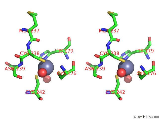
Zinc binding site 1 out of 4 in 4kvp
Go back to
Zinc binding site 1 out
of 4 in the Human P53 Core Domain Mutant V157F
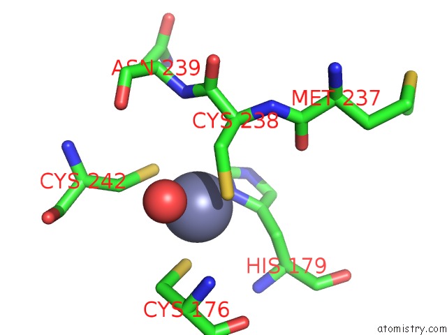
Mono view
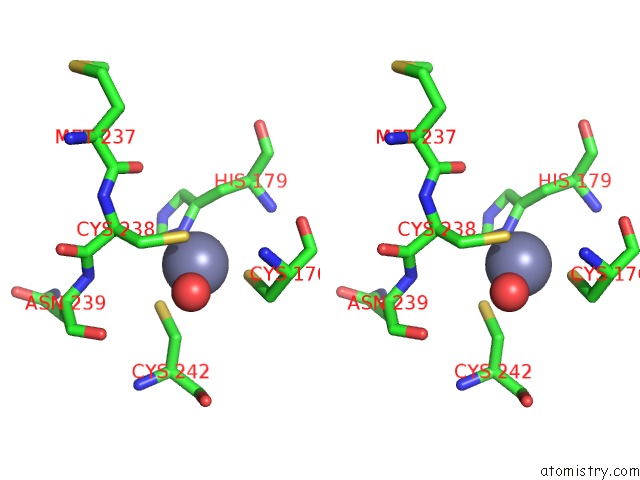
Stereo pair view
Mono view
Stereo pair view
A full contact list of Zinc with other atoms in the Zn binding
site number 1 of Human P53 Core Domain Mutant V157F within 5.0Å range:
|
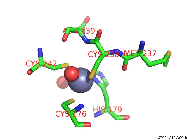
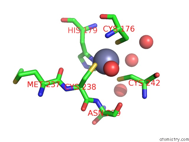
Zinc binding site 2 out of 4 in 4kvp
Go back to
Zinc binding site 2 out
of 4 in the Human P53 Core Domain Mutant V157F

Mono view
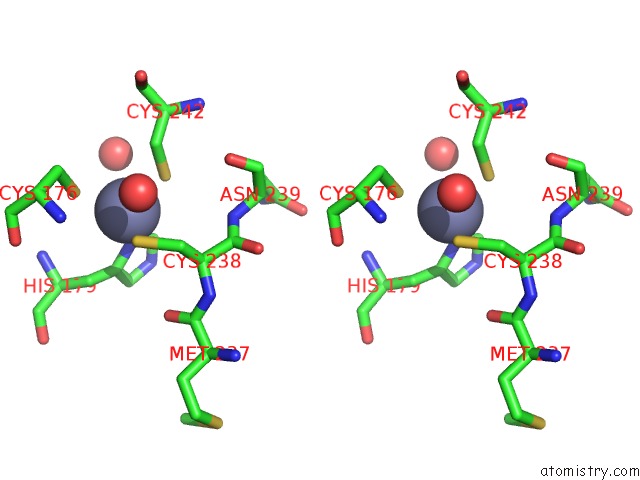
Stereo pair view
Mono view
Stereo pair view
A full contact list of Zinc with other atoms in the Zn binding
site number 2 of Human P53 Core Domain Mutant V157F within 5.0Å range:
|
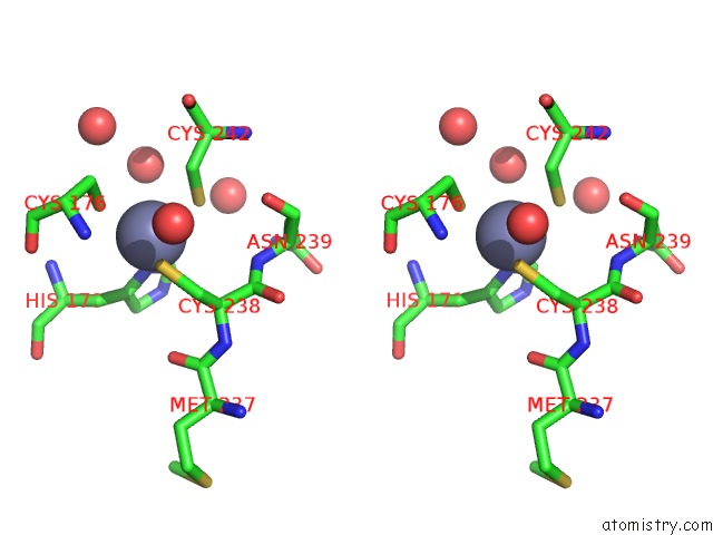
Zinc binding site 3 out of 4 in 4kvp
Go back to
Zinc binding site 3 out
of 4 in the Human P53 Core Domain Mutant V157F
Mono view
Stereo pair view
Mono view
Stereo pair view
A full contact list of Zinc with other atoms in the Zn binding
site number 3 of Human P53 Core Domain Mutant V157F within 5.0Å range:
|
Zinc binding site 4 out of 4 in 4kvp
Go back to
Zinc binding site 4 out
of 4 in the Human P53 Core Domain Mutant V157F
Mono view
Stereo pair view
Mono view
Stereo pair view
A full contact list of Zinc with other atoms in the Zn binding
site number 4 of Human P53 Core Domain Mutant V157F within 5.0Å range:
|
Reference:
B.D.Wallentine,
Y.Wang,
V.Tretyachenko-Ladokhina,
M.Tan,
D.F.Senear,
H.Luecke.
Structures of Oncogenic, Suppressor and Rescued P53 Core-Domain Variants: Mechanisms of Mutant P53 Rescue. Acta Crystallogr.,Sect.D V. 69 2146 2013.
ISSN: ISSN 0907-4449
PubMed: 24100332
DOI: 10.1107/S0907444913020830
Page generated: Sun Oct 27 01:23:24 2024
ISSN: ISSN 0907-4449
PubMed: 24100332
DOI: 10.1107/S0907444913020830
Last articles
Zn in 9MJ5Zn in 9HNW
Zn in 9G0L
Zn in 9FNE
Zn in 9DZN
Zn in 9E0I
Zn in 9D32
Zn in 9DAK
Zn in 8ZXC
Zn in 8ZUF