Zinc »
PDB 4kjv-4kxq »
4koy »
Zinc in PDB 4koy: Crystal Structure of A Gnat Superfamily Acetyltransferase PA4794 in Complex with Cephalosporin C
Protein crystallography data
The structure of Crystal Structure of A Gnat Superfamily Acetyltransferase PA4794 in Complex with Cephalosporin C, PDB code: 4koy
was solved by
K.A.Majorek,
P.J.Porebski,
M.Chruszcz,
M.Cymborowski,
A.Joachimiak,
W.Minor,
Midwest Center For Structural Genomics (Mcsg),
with X-Ray Crystallography technique. A brief refinement statistics is given in the table below:
| Resolution Low / High (Å) | 34.94 / 1.40 |
| Space group | P 21 21 2 |
| Cell size a, b, c (Å), α, β, γ (°) | 58.000, 76.059, 39.303, 90.00, 90.00, 90.00 |
| R / Rfree (%) | 14.6 / 17.3 |
Zinc Binding Sites:
The binding sites of Zinc atom in the Crystal Structure of A Gnat Superfamily Acetyltransferase PA4794 in Complex with Cephalosporin C
(pdb code 4koy). This binding sites where shown within
5.0 Angstroms radius around Zinc atom.
In total 4 binding sites of Zinc where determined in the Crystal Structure of A Gnat Superfamily Acetyltransferase PA4794 in Complex with Cephalosporin C, PDB code: 4koy:
Jump to Zinc binding site number: 1; 2; 3; 4;
In total 4 binding sites of Zinc where determined in the Crystal Structure of A Gnat Superfamily Acetyltransferase PA4794 in Complex with Cephalosporin C, PDB code: 4koy:
Jump to Zinc binding site number: 1; 2; 3; 4;
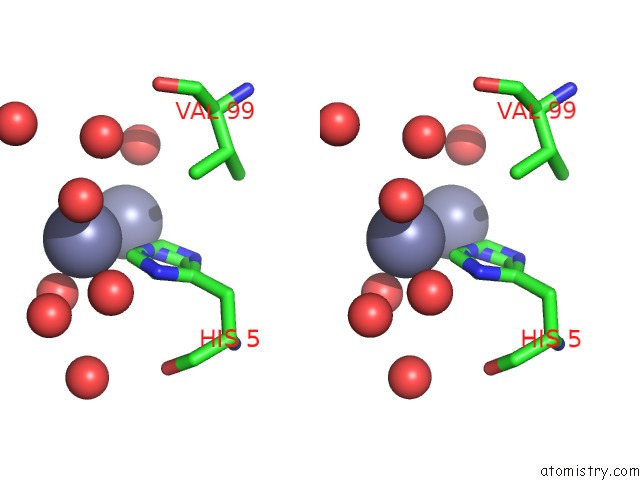
Zinc binding site 1 out of 4 in 4koy
Go back to
Zinc binding site 1 out
of 4 in the Crystal Structure of A Gnat Superfamily Acetyltransferase PA4794 in Complex with Cephalosporin C

Mono view
Stereo pair view
Mono view
Stereo pair view
A full contact list of Zinc with other atoms in the Zn binding
site number 1 of Crystal Structure of A Gnat Superfamily Acetyltransferase PA4794 in Complex with Cephalosporin C within 5.0Å range:
|
Zinc binding site 2 out of 4 in 4koy
Go back to
Zinc binding site 2 out
of 4 in the Crystal Structure of A Gnat Superfamily Acetyltransferase PA4794 in Complex with Cephalosporin C

Mono view

Stereo pair view
Mono view
Stereo pair view
A full contact list of Zinc with other atoms in the Zn binding
site number 2 of Crystal Structure of A Gnat Superfamily Acetyltransferase PA4794 in Complex with Cephalosporin C within 5.0Å range:
|
Zinc binding site 3 out of 4 in 4koy
Go back to
Zinc binding site 3 out
of 4 in the Crystal Structure of A Gnat Superfamily Acetyltransferase PA4794 in Complex with Cephalosporin C

Mono view

Stereo pair view
Mono view
Stereo pair view
A full contact list of Zinc with other atoms in the Zn binding
site number 3 of Crystal Structure of A Gnat Superfamily Acetyltransferase PA4794 in Complex with Cephalosporin C within 5.0Å range:
|
Zinc binding site 4 out of 4 in 4koy
Go back to
Zinc binding site 4 out
of 4 in the Crystal Structure of A Gnat Superfamily Acetyltransferase PA4794 in Complex with Cephalosporin C

Mono view

Stereo pair view
Mono view
Stereo pair view
A full contact list of Zinc with other atoms in the Zn binding
site number 4 of Crystal Structure of A Gnat Superfamily Acetyltransferase PA4794 in Complex with Cephalosporin C within 5.0Å range:
|
Reference:
K.A.Majorek,
M.L.Kuhn,
M.Chruszcz,
W.F.Anderson,
W.Minor.
Structural, Functional, and Inhibition Studies of A GCN5-Related N-Acetyltransferase (Gnat) Superfamily Protein PA4794: A New C-Terminal Lysine Protein Acetyltransferase From Pseudomonas Aeruginosa. J.Biol.Chem. V. 288 30223 2013.
ISSN: ISSN 0021-9258
PubMed: 24003232
DOI: 10.1074/JBC.M113.501353
Page generated: Sun Oct 27 01:20:09 2024
ISSN: ISSN 0021-9258
PubMed: 24003232
DOI: 10.1074/JBC.M113.501353
Last articles
Zn in 9MJ5Zn in 9HNW
Zn in 9G0L
Zn in 9FNE
Zn in 9DZN
Zn in 9E0I
Zn in 9D32
Zn in 9DAK
Zn in 8ZXC
Zn in 8ZUF