Zinc »
PDB 4h9d-4hki »
4hdt »
Zinc in PDB 4hdt: Crystal Structure of A Carnitinyl-Coa Dehydratase From Mycobacterium Thermoresistibile
Protein crystallography data
The structure of Crystal Structure of A Carnitinyl-Coa Dehydratase From Mycobacterium Thermoresistibile, PDB code: 4hdt
was solved by
Seattle Structural Genomics Center For Infectious Disease (Ssgcid),
with X-Ray Crystallography technique. A brief refinement statistics is given in the table below:
| Resolution Low / High (Å) | 45.45 / 1.60 |
| Space group | P 1 21 1 |
| Cell size a, b, c (Å), α, β, γ (°) | 48.200, 62.400, 56.880, 90.00, 109.59, 90.00 |
| R / Rfree (%) | 16 / 18.9 |
Other elements in 4hdt:
The structure of Crystal Structure of A Carnitinyl-Coa Dehydratase From Mycobacterium Thermoresistibile also contains other interesting chemical elements:
| Chlorine | (Cl) | 1 atom |
Zinc Binding Sites:
The binding sites of Zinc atom in the Crystal Structure of A Carnitinyl-Coa Dehydratase From Mycobacterium Thermoresistibile
(pdb code 4hdt). This binding sites where shown within
5.0 Angstroms radius around Zinc atom.
In total 5 binding sites of Zinc where determined in the Crystal Structure of A Carnitinyl-Coa Dehydratase From Mycobacterium Thermoresistibile, PDB code: 4hdt:
Jump to Zinc binding site number: 1; 2; 3; 4; 5;
In total 5 binding sites of Zinc where determined in the Crystal Structure of A Carnitinyl-Coa Dehydratase From Mycobacterium Thermoresistibile, PDB code: 4hdt:
Jump to Zinc binding site number: 1; 2; 3; 4; 5;
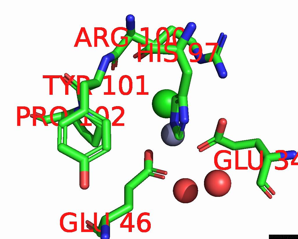
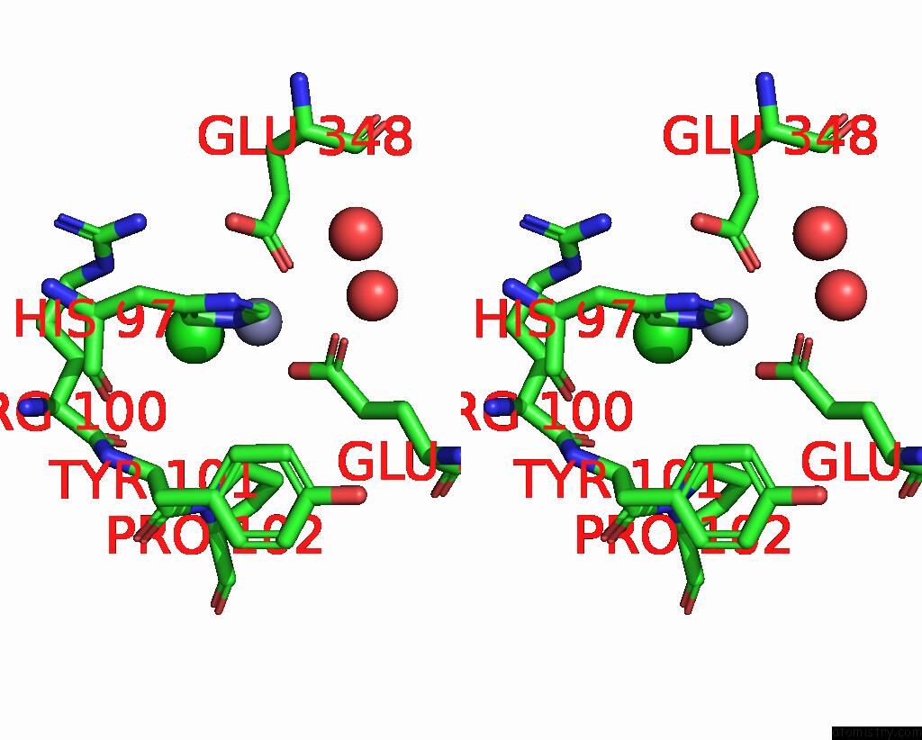
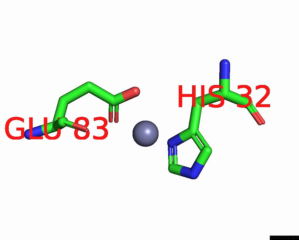
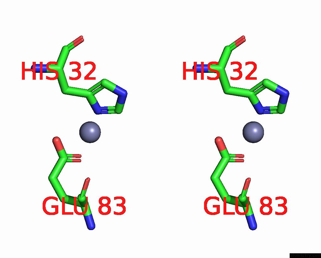
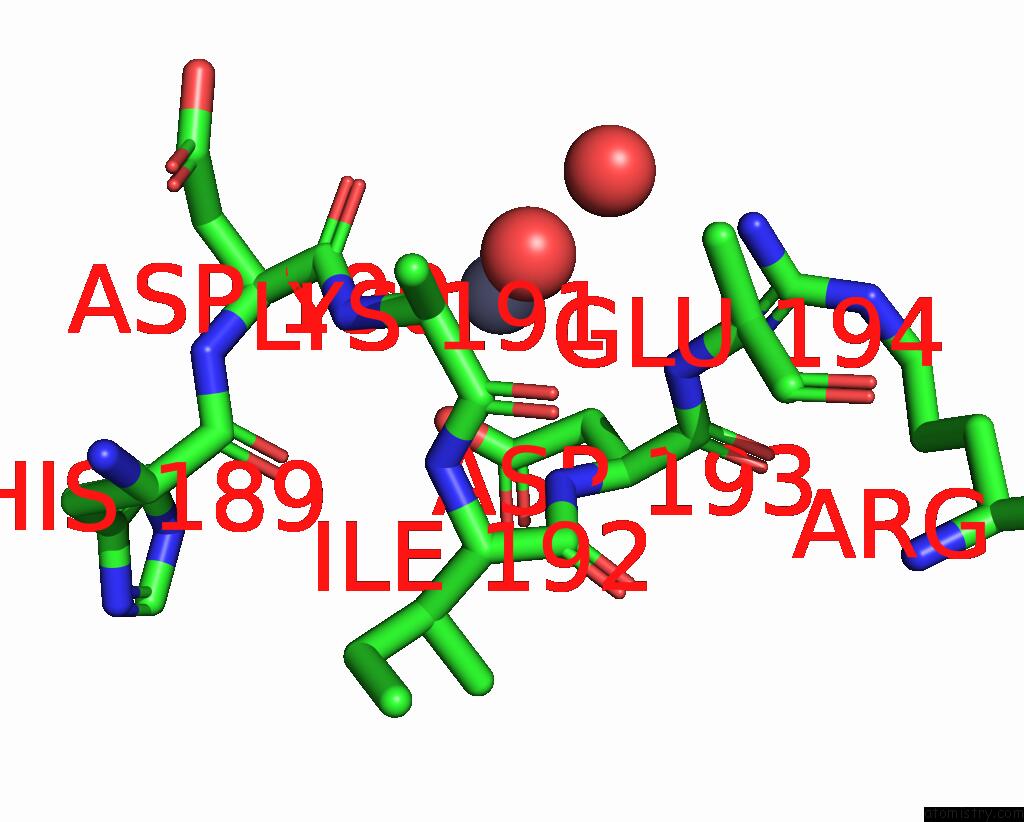
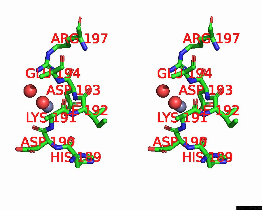
Zinc binding site 1 out of 5 in 4hdt
Go back to
Zinc binding site 1 out
of 5 in the Crystal Structure of A Carnitinyl-Coa Dehydratase From Mycobacterium Thermoresistibile
Mono view
Stereo pair view
Mono view
Stereo pair view
A full contact list of Zinc with other atoms in the Zn binding
site number 1 of Crystal Structure of A Carnitinyl-Coa Dehydratase From Mycobacterium Thermoresistibile within 5.0Å range:
|
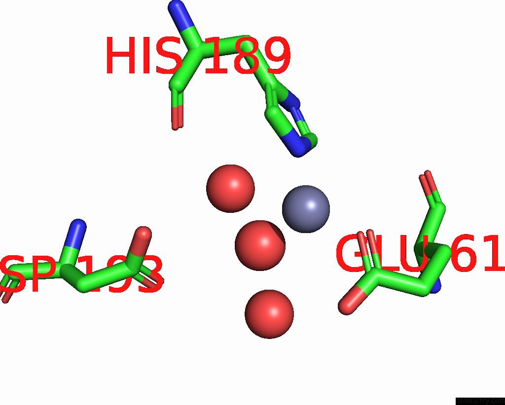
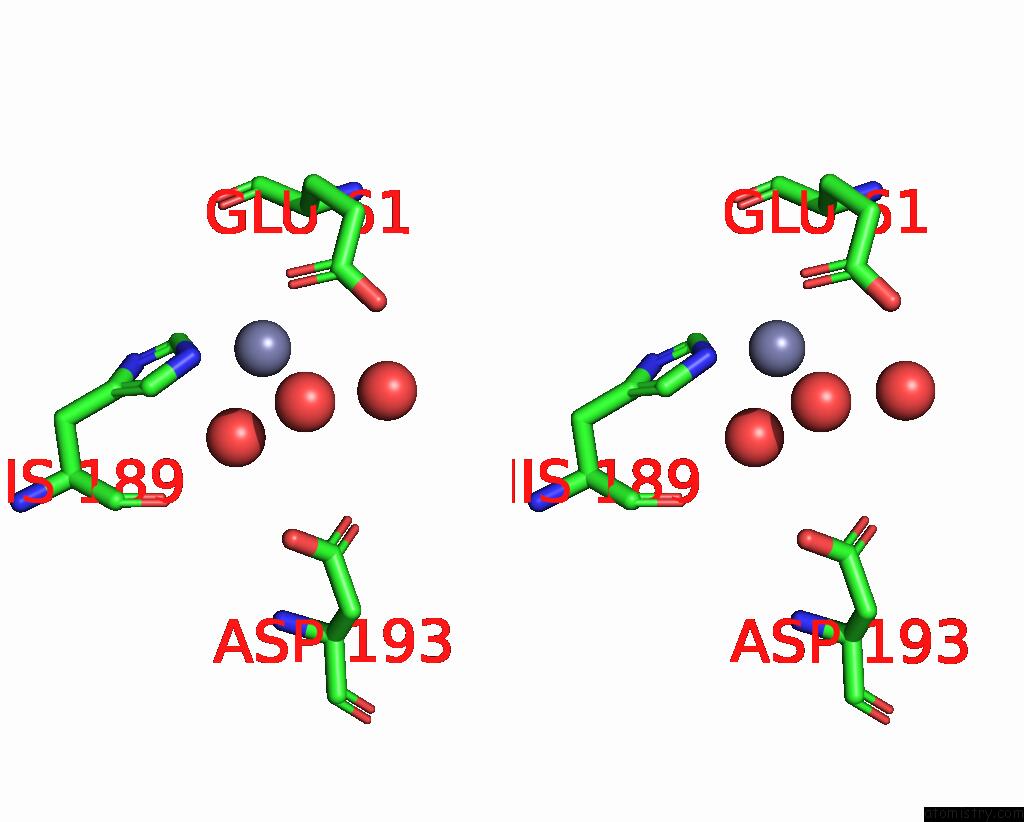
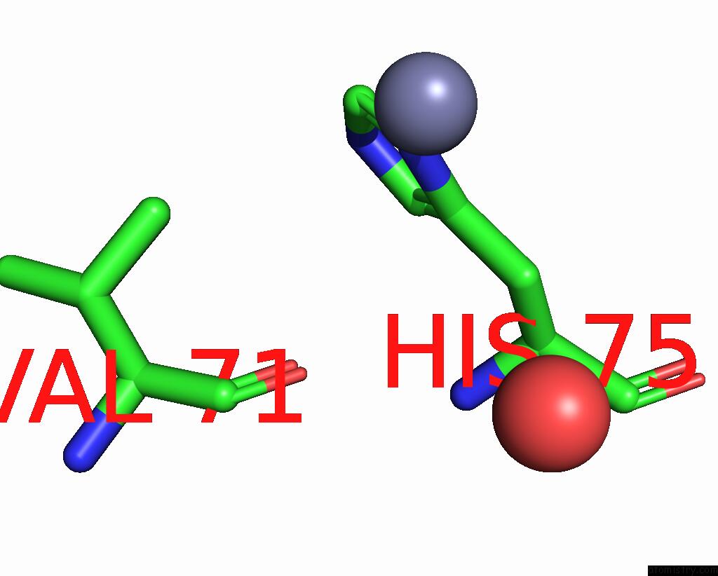
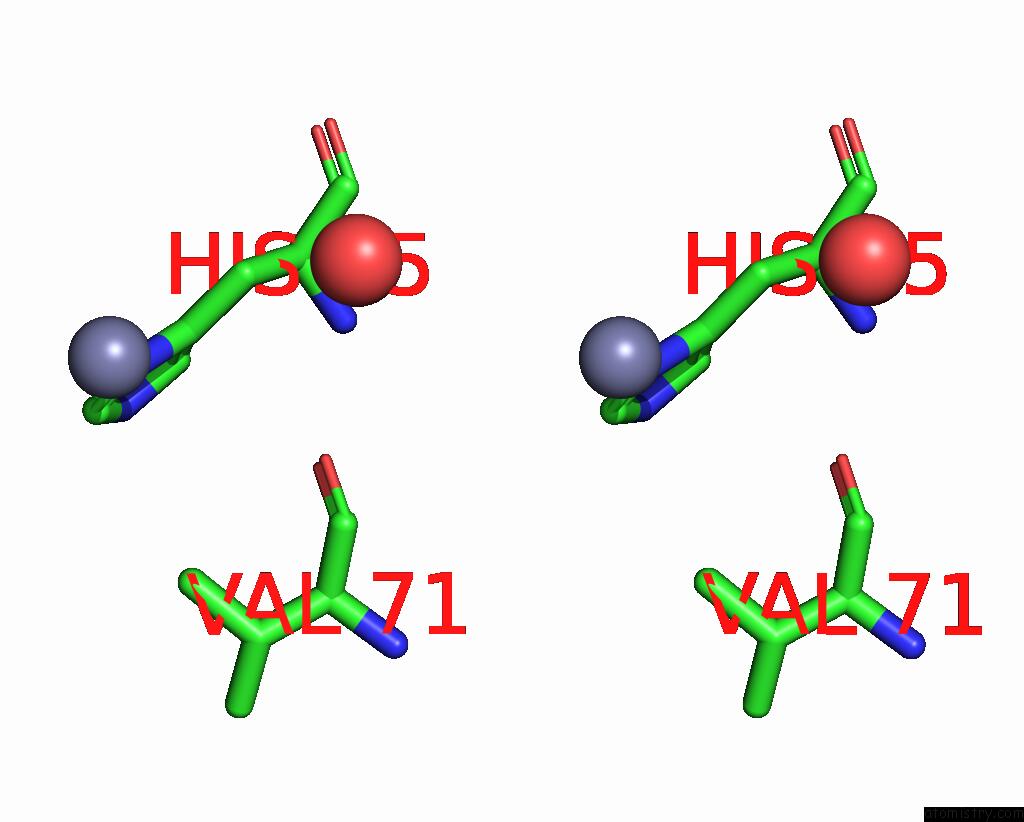
Zinc binding site 2 out of 5 in 4hdt
Go back to
Zinc binding site 2 out
of 5 in the Crystal Structure of A Carnitinyl-Coa Dehydratase From Mycobacterium Thermoresistibile
Mono view
Stereo pair view
Mono view
Stereo pair view
A full contact list of Zinc with other atoms in the Zn binding
site number 2 of Crystal Structure of A Carnitinyl-Coa Dehydratase From Mycobacterium Thermoresistibile within 5.0Å range:
|
Zinc binding site 3 out of 5 in 4hdt
Go back to
Zinc binding site 3 out
of 5 in the Crystal Structure of A Carnitinyl-Coa Dehydratase From Mycobacterium Thermoresistibile
Mono view
Stereo pair view
Mono view
Stereo pair view
A full contact list of Zinc with other atoms in the Zn binding
site number 3 of Crystal Structure of A Carnitinyl-Coa Dehydratase From Mycobacterium Thermoresistibile within 5.0Å range:
|
Zinc binding site 4 out of 5 in 4hdt
Go back to
Zinc binding site 4 out
of 5 in the Crystal Structure of A Carnitinyl-Coa Dehydratase From Mycobacterium Thermoresistibile
Mono view
Stereo pair view
Mono view
Stereo pair view
A full contact list of Zinc with other atoms in the Zn binding
site number 4 of Crystal Structure of A Carnitinyl-Coa Dehydratase From Mycobacterium Thermoresistibile within 5.0Å range:
|
Zinc binding site 5 out of 5 in 4hdt
Go back to
Zinc binding site 5 out
of 5 in the Crystal Structure of A Carnitinyl-Coa Dehydratase From Mycobacterium Thermoresistibile
Mono view
Stereo pair view
Mono view
Stereo pair view
A full contact list of Zinc with other atoms in the Zn binding
site number 5 of Crystal Structure of A Carnitinyl-Coa Dehydratase From Mycobacterium Thermoresistibile within 5.0Å range:
|
Reference:
L.Baugh,
I.Phan,
D.W.Begley,
M.C.Clifton,
B.Armour,
D.M.Dranow,
B.M.Taylor,
M.M.Muruthi,
J.Abendroth,
J.W.Fairman,
D.Fox,
S.H.Dieterich,
B.L.Staker,
A.S.Gardberg,
R.Choi,
S.N.Hewitt,
A.J.Napuli,
J.Myers,
L.K.Barrett,
Y.Zhang,
M.Ferrell,
E.Mundt,
K.Thompkins,
N.Tran,
S.Lyons-Abbott,
A.Abramov,
A.Sekar,
D.Serbzhinskiy,
D.Lorimer,
G.W.Buchko,
R.Stacy,
L.J.Stewart,
T.E.Edwards,
W.C.Van Voorhis,
P.J.Myler.
Increasing the Structural Coverage of Tuberculosis Drug Targets. Tuberculosis (Edinb) V. 95 142 2015.
ISSN: ISSN 1472-9792
PubMed: 25613812
DOI: 10.1016/J.TUBE.2014.12.003
Page generated: Sun Oct 27 00:02:13 2024
ISSN: ISSN 1472-9792
PubMed: 25613812
DOI: 10.1016/J.TUBE.2014.12.003
Last articles
Zn in 9MJ5Zn in 9HNW
Zn in 9G0L
Zn in 9FNE
Zn in 9DZN
Zn in 9E0I
Zn in 9D32
Zn in 9DAK
Zn in 8ZXC
Zn in 8ZUF