Zinc »
PDB 4f78-4fke »
4fil »
Zinc in PDB 4fil: Structure of FHUD2 From Staphylococcus Aureus with Bound Ferrioxamine B
Protein crystallography data
The structure of Structure of FHUD2 From Staphylococcus Aureus with Bound Ferrioxamine B, PDB code: 4fil
was solved by
L.K.Briere,
D.E.Heinrichs,
B.H.Shilton,
with X-Ray Crystallography technique. A brief refinement statistics is given in the table below:
| Resolution Low / High (Å) | 45.00 / 2.40 |
| Space group | P 1 21 1 |
| Cell size a, b, c (Å), α, β, γ (°) | 81.343, 78.996, 116.811, 90.00, 109.87, 90.00 |
| R / Rfree (%) | 19.8 / 22.9 |
Other elements in 4fil:
The structure of Structure of FHUD2 From Staphylococcus Aureus with Bound Ferrioxamine B also contains other interesting chemical elements:
| Iron | (Fe) | 4 atoms |
Zinc Binding Sites:
Pages:
>>> Page 1 <<< Page 2, Binding sites: 11 - 20; Page 3, Binding sites: 21 - 30; Page 4, Binding sites: 31 - 40; Page 5, Binding sites: 41 - 50; Page 6, Binding sites: 51 - 56;Binding sites:
The binding sites of Zinc atom in the Structure of FHUD2 From Staphylococcus Aureus with Bound Ferrioxamine B (pdb code 4fil). This binding sites where shown within 5.0 Angstroms radius around Zinc atom.In total 56 binding sites of Zinc where determined in the Structure of FHUD2 From Staphylococcus Aureus with Bound Ferrioxamine B, PDB code: 4fil:
Jump to Zinc binding site number: 1; 2; 3; 4; 5; 6; 7; 8; 9; 10;
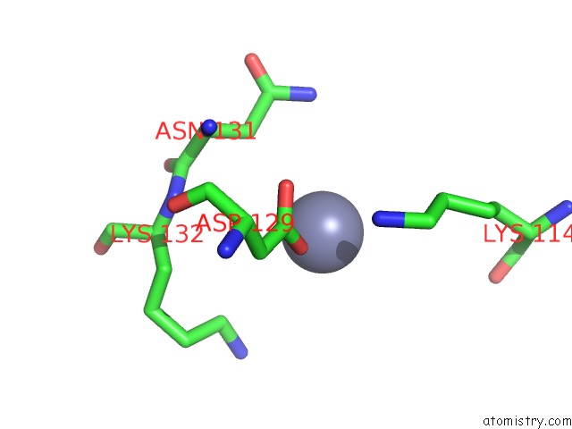
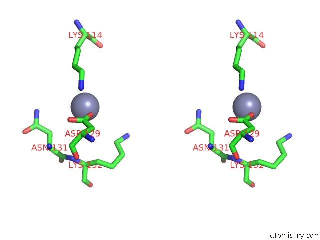
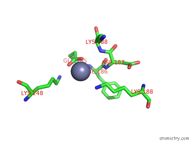
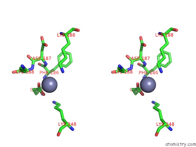
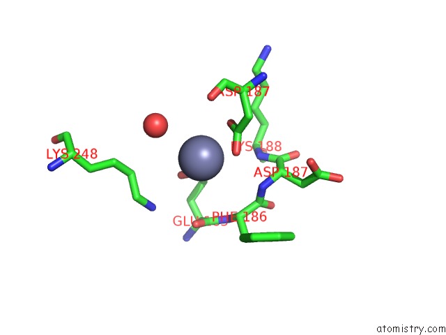
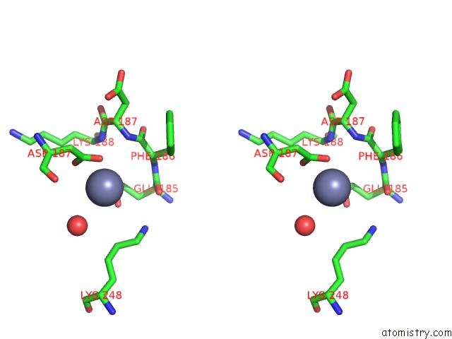
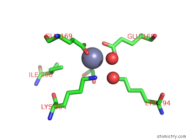
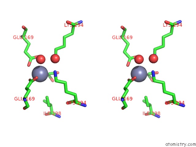
Zinc binding site 1 out of 56 in 4fil
Go back to
Zinc binding site 1 out
of 56 in the Structure of FHUD2 From Staphylococcus Aureus with Bound Ferrioxamine B
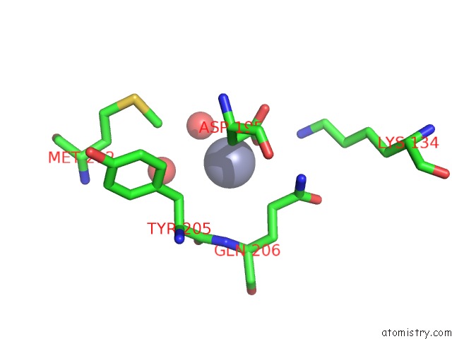
Mono view
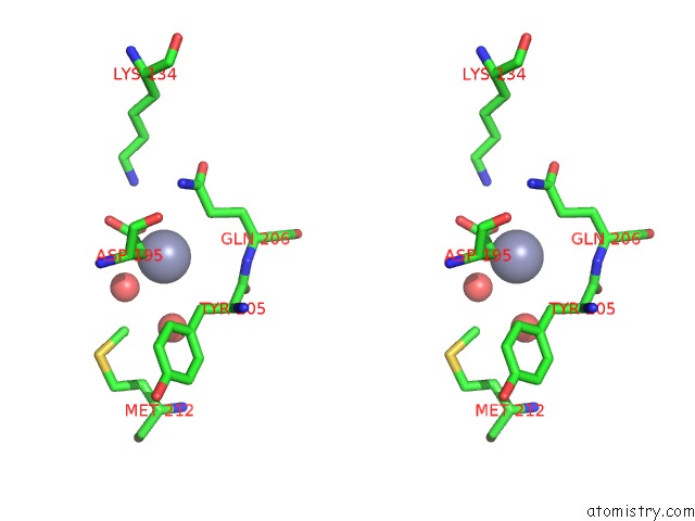
Stereo pair view
Mono view
Stereo pair view
A full contact list of Zinc with other atoms in the Zn binding
site number 1 of Structure of FHUD2 From Staphylococcus Aureus with Bound Ferrioxamine B within 5.0Å range:
|
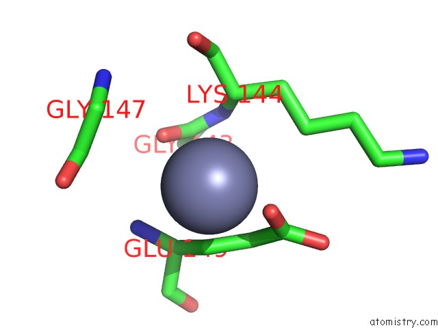
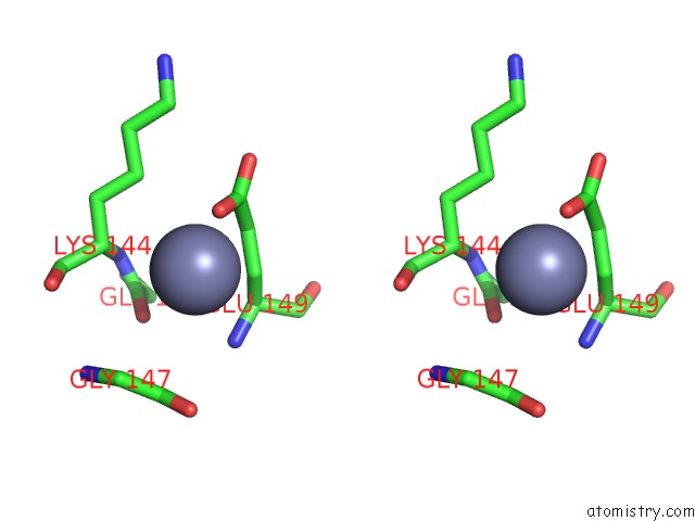
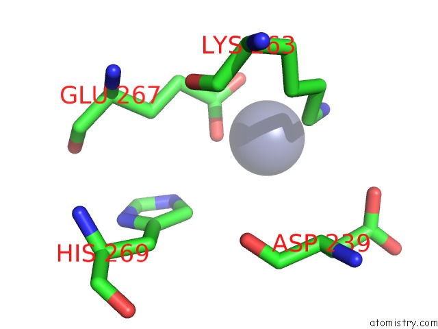
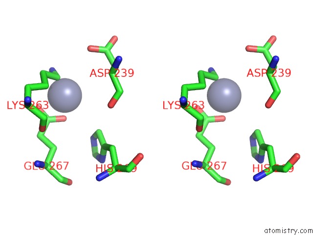
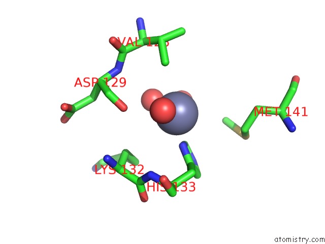
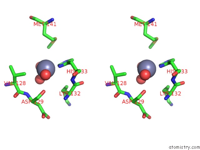
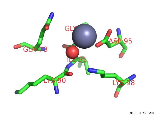
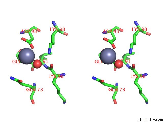
Zinc binding site 2 out of 56 in 4fil
Go back to
Zinc binding site 2 out
of 56 in the Structure of FHUD2 From Staphylococcus Aureus with Bound Ferrioxamine B
Mono view
Stereo pair view
Mono view
Stereo pair view
A full contact list of Zinc with other atoms in the Zn binding
site number 2 of Structure of FHUD2 From Staphylococcus Aureus with Bound Ferrioxamine B within 5.0Å range:
|
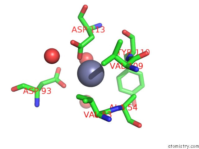
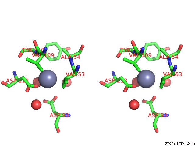
Zinc binding site 3 out of 56 in 4fil
Go back to
Zinc binding site 3 out
of 56 in the Structure of FHUD2 From Staphylococcus Aureus with Bound Ferrioxamine B
Mono view
Stereo pair view
Mono view
Stereo pair view
A full contact list of Zinc with other atoms in the Zn binding
site number 3 of Structure of FHUD2 From Staphylococcus Aureus with Bound Ferrioxamine B within 5.0Å range:
|
Zinc binding site 4 out of 56 in 4fil
Go back to
Zinc binding site 4 out
of 56 in the Structure of FHUD2 From Staphylococcus Aureus with Bound Ferrioxamine B
Mono view
Stereo pair view
Mono view
Stereo pair view
A full contact list of Zinc with other atoms in the Zn binding
site number 4 of Structure of FHUD2 From Staphylococcus Aureus with Bound Ferrioxamine B within 5.0Å range:
|
Zinc binding site 5 out of 56 in 4fil
Go back to
Zinc binding site 5 out
of 56 in the Structure of FHUD2 From Staphylococcus Aureus with Bound Ferrioxamine B
Mono view
Stereo pair view
Mono view
Stereo pair view
A full contact list of Zinc with other atoms in the Zn binding
site number 5 of Structure of FHUD2 From Staphylococcus Aureus with Bound Ferrioxamine B within 5.0Å range:
|
Zinc binding site 6 out of 56 in 4fil
Go back to
Zinc binding site 6 out
of 56 in the Structure of FHUD2 From Staphylococcus Aureus with Bound Ferrioxamine B
Mono view
Stereo pair view
Mono view
Stereo pair view
A full contact list of Zinc with other atoms in the Zn binding
site number 6 of Structure of FHUD2 From Staphylococcus Aureus with Bound Ferrioxamine B within 5.0Å range:
|
Zinc binding site 7 out of 56 in 4fil
Go back to
Zinc binding site 7 out
of 56 in the Structure of FHUD2 From Staphylococcus Aureus with Bound Ferrioxamine B
Mono view
Stereo pair view
Mono view
Stereo pair view
A full contact list of Zinc with other atoms in the Zn binding
site number 7 of Structure of FHUD2 From Staphylococcus Aureus with Bound Ferrioxamine B within 5.0Å range:
|
Zinc binding site 8 out of 56 in 4fil
Go back to
Zinc binding site 8 out
of 56 in the Structure of FHUD2 From Staphylococcus Aureus with Bound Ferrioxamine B
Mono view
Stereo pair view
Mono view
Stereo pair view
A full contact list of Zinc with other atoms in the Zn binding
site number 8 of Structure of FHUD2 From Staphylococcus Aureus with Bound Ferrioxamine B within 5.0Å range:
|
Zinc binding site 9 out of 56 in 4fil
Go back to
Zinc binding site 9 out
of 56 in the Structure of FHUD2 From Staphylococcus Aureus with Bound Ferrioxamine B
Mono view
Stereo pair view
Mono view
Stereo pair view
A full contact list of Zinc with other atoms in the Zn binding
site number 9 of Structure of FHUD2 From Staphylococcus Aureus with Bound Ferrioxamine B within 5.0Å range:
|
Zinc binding site 10 out of 56 in 4fil
Go back to
Zinc binding site 10 out
of 56 in the Structure of FHUD2 From Staphylococcus Aureus with Bound Ferrioxamine B
Mono view
Stereo pair view
Mono view
Stereo pair view
A full contact list of Zinc with other atoms in the Zn binding
site number 10 of Structure of FHUD2 From Staphylococcus Aureus with Bound Ferrioxamine B within 5.0Å range:
|
Reference:
K.J.Podkowa,
L.A.Briere,
D.E.Heinrichs,
B.H.Shilton.
Crystal and Solution Structure Analysis of FHUD2 From Staphylococcus Aureus in Multiple Unliganded Conformations and Bound to Ferrioxamine-B. Biochemistry V. 53 2017 2014.
ISSN: ISSN 0006-2960
PubMed: 24606332
DOI: 10.1021/BI401349D
Page generated: Sat Oct 26 22:28:09 2024
ISSN: ISSN 0006-2960
PubMed: 24606332
DOI: 10.1021/BI401349D
Last articles
Zn in 9MJ5Zn in 9HNW
Zn in 9G0L
Zn in 9FNE
Zn in 9DZN
Zn in 9E0I
Zn in 9D32
Zn in 9DAK
Zn in 8ZXC
Zn in 8ZUF