Zinc »
PDB 4csz-4cyk »
4cvt »
Zinc in PDB 4cvt: Structure of Apobacterioferritin Y58F Variant
Protein crystallography data
The structure of Structure of Apobacterioferritin Y58F Variant, PDB code: 4cvt
was solved by
K.Hingorani,
R.Pace,
S.Whitney,
J.W.Murray,
T.Wydrzynski,
M.H.Cheah,
P.Smith,
W.Hillier,
with X-Ray Crystallography technique. A brief refinement statistics is given in the table below:
| Resolution Low / High (Å) | 46.817 / 1.79 |
| Space group | C 1 2 1 |
| Cell size a, b, c (Å), α, β, γ (°) | 104.460, 28.070, 56.810, 90.00, 119.18, 90.00 |
| R / Rfree (%) | 16.72 / 21.42 |
Zinc Binding Sites:
The binding sites of Zinc atom in the Structure of Apobacterioferritin Y58F Variant
(pdb code 4cvt). This binding sites where shown within
5.0 Angstroms radius around Zinc atom.
In total 4 binding sites of Zinc where determined in the Structure of Apobacterioferritin Y58F Variant, PDB code: 4cvt:
Jump to Zinc binding site number: 1; 2; 3; 4;
In total 4 binding sites of Zinc where determined in the Structure of Apobacterioferritin Y58F Variant, PDB code: 4cvt:
Jump to Zinc binding site number: 1; 2; 3; 4;
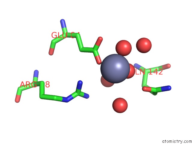
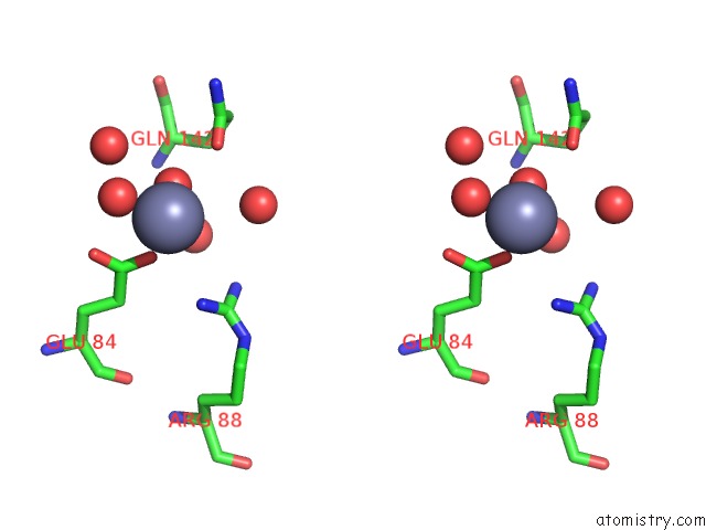
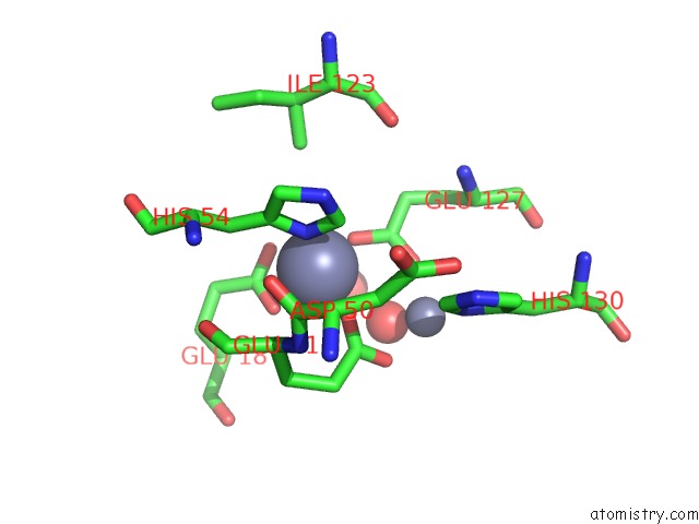
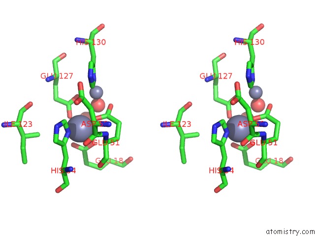
Zinc binding site 1 out of 4 in 4cvt
Go back to
Zinc binding site 1 out
of 4 in the Structure of Apobacterioferritin Y58F Variant
Mono view
Stereo pair view
Mono view
Stereo pair view
A full contact list of Zinc with other atoms in the Zn binding
site number 1 of Structure of Apobacterioferritin Y58F Variant within 5.0Å range:
|
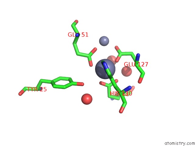
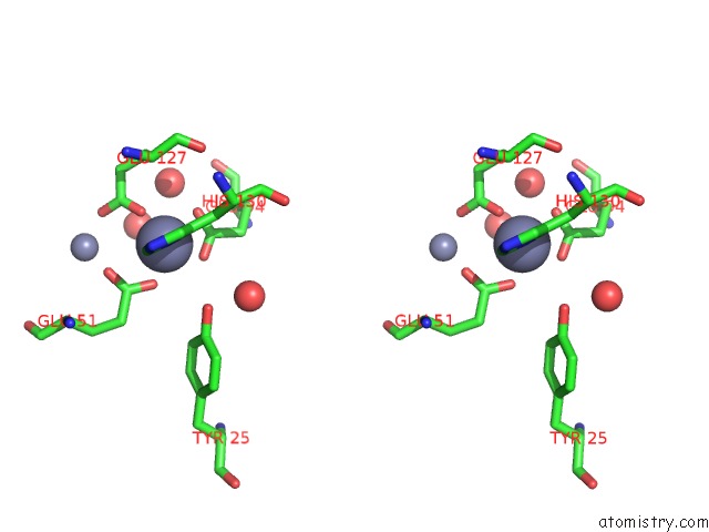
Zinc binding site 2 out of 4 in 4cvt
Go back to
Zinc binding site 2 out
of 4 in the Structure of Apobacterioferritin Y58F Variant
Mono view
Stereo pair view
Mono view
Stereo pair view
A full contact list of Zinc with other atoms in the Zn binding
site number 2 of Structure of Apobacterioferritin Y58F Variant within 5.0Å range:
|
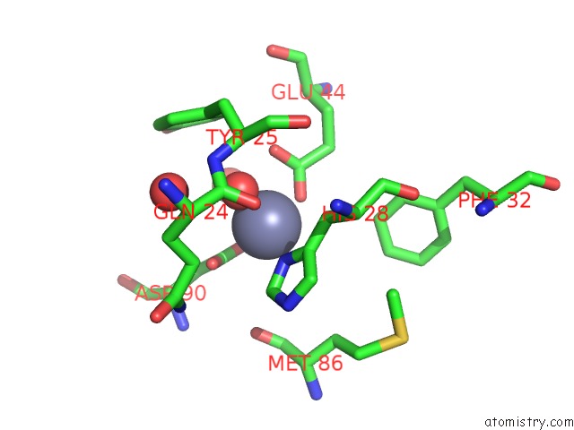
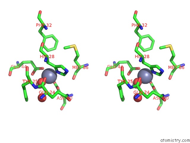
Zinc binding site 3 out of 4 in 4cvt
Go back to
Zinc binding site 3 out
of 4 in the Structure of Apobacterioferritin Y58F Variant
Mono view
Stereo pair view
Mono view
Stereo pair view
A full contact list of Zinc with other atoms in the Zn binding
site number 3 of Structure of Apobacterioferritin Y58F Variant within 5.0Å range:
|
Zinc binding site 4 out of 4 in 4cvt
Go back to
Zinc binding site 4 out
of 4 in the Structure of Apobacterioferritin Y58F Variant
Mono view
Stereo pair view
Mono view
Stereo pair view
A full contact list of Zinc with other atoms in the Zn binding
site number 4 of Structure of Apobacterioferritin Y58F Variant within 5.0Å range:
|
Reference:
K.Hingorani,
R.Pace,
S.Whitney,
J.W.Murray,
P.Smith,
M.H.Cheah,
T.Wydrzynski,
W.Hillier.
Photo-Oxidation of Tyrosine in A Bio-Engineered Bacterioferritin 'Reaction Centre'-A Protein Model For Artificial Photosynthesis. Biochim.Biophys.Acta V.1837 1821 2014.
ISSN: ISSN 0006-3002
PubMed: 25107631
DOI: 10.1016/J.BBABIO.2014.07.019
Page generated: Sat Oct 26 21:07:29 2024
ISSN: ISSN 0006-3002
PubMed: 25107631
DOI: 10.1016/J.BBABIO.2014.07.019
Last articles
Zn in 9MJ5Zn in 9HNW
Zn in 9G0L
Zn in 9FNE
Zn in 9DZN
Zn in 9E0I
Zn in 9D32
Zn in 9DAK
Zn in 8ZXC
Zn in 8ZUF