Zinc »
PDB 4cis-4csp »
4cs7 »
Zinc in PDB 4cs7: Crystal Structure of the Asymmetric Human Metapneumovirus M2-1 Tetramer, Form 1
Protein crystallography data
The structure of Crystal Structure of the Asymmetric Human Metapneumovirus M2-1 Tetramer, Form 1, PDB code: 4cs7
was solved by
C.Leyrat,
M.Renner,
K.Harlos,
J.M.Grimes,
with X-Ray Crystallography technique. A brief refinement statistics is given in the table below:
| Resolution Low / High (Å) | 33.13 / 2.47 |
| Space group | P 1 21 1 |
| Cell size a, b, c (Å), α, β, γ (°) | 50.150, 92.670, 82.830, 90.00, 94.48, 90.00 |
| R / Rfree (%) | 23.34 / 26.15 |
Zinc Binding Sites:
The binding sites of Zinc atom in the Crystal Structure of the Asymmetric Human Metapneumovirus M2-1 Tetramer, Form 1
(pdb code 4cs7). This binding sites where shown within
5.0 Angstroms radius around Zinc atom.
In total 4 binding sites of Zinc where determined in the Crystal Structure of the Asymmetric Human Metapneumovirus M2-1 Tetramer, Form 1, PDB code: 4cs7:
Jump to Zinc binding site number: 1; 2; 3; 4;
In total 4 binding sites of Zinc where determined in the Crystal Structure of the Asymmetric Human Metapneumovirus M2-1 Tetramer, Form 1, PDB code: 4cs7:
Jump to Zinc binding site number: 1; 2; 3; 4;
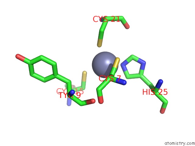
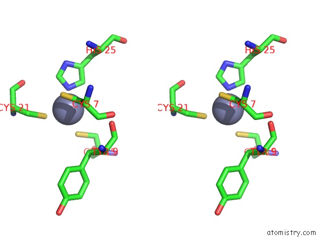
Zinc binding site 1 out of 4 in 4cs7
Go back to
Zinc binding site 1 out
of 4 in the Crystal Structure of the Asymmetric Human Metapneumovirus M2-1 Tetramer, Form 1
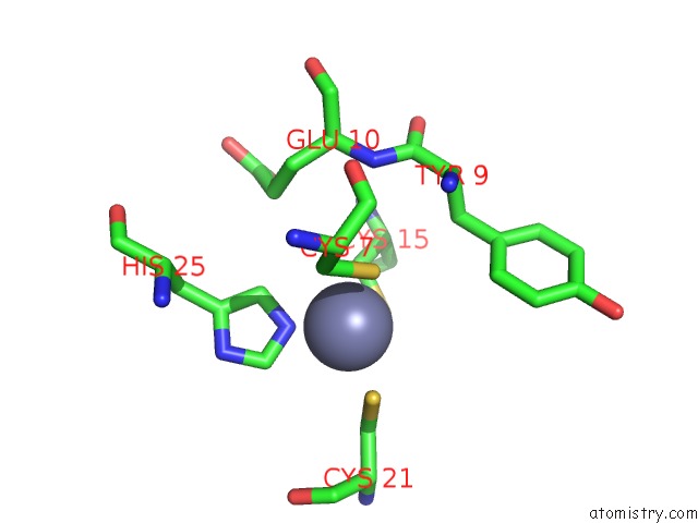
Mono view
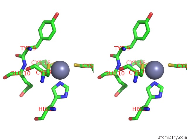
Stereo pair view
Mono view
Stereo pair view
A full contact list of Zinc with other atoms in the Zn binding
site number 1 of Crystal Structure of the Asymmetric Human Metapneumovirus M2-1 Tetramer, Form 1 within 5.0Å range:
|
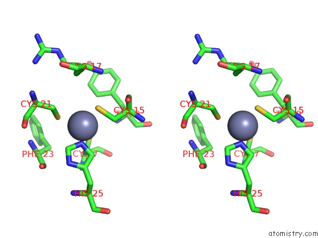
Zinc binding site 2 out of 4 in 4cs7
Go back to
Zinc binding site 2 out
of 4 in the Crystal Structure of the Asymmetric Human Metapneumovirus M2-1 Tetramer, Form 1
Mono view
Stereo pair view
Mono view
Stereo pair view
A full contact list of Zinc with other atoms in the Zn binding
site number 2 of Crystal Structure of the Asymmetric Human Metapneumovirus M2-1 Tetramer, Form 1 within 5.0Å range:
|
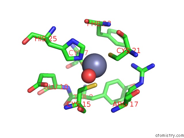
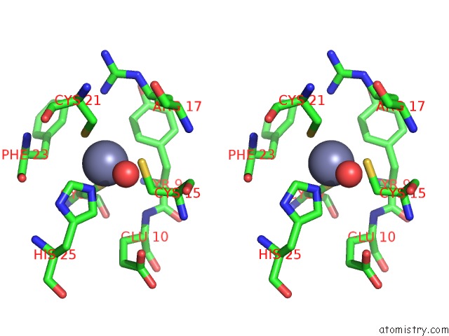
Zinc binding site 3 out of 4 in 4cs7
Go back to
Zinc binding site 3 out
of 4 in the Crystal Structure of the Asymmetric Human Metapneumovirus M2-1 Tetramer, Form 1
Mono view
Stereo pair view
Mono view
Stereo pair view
A full contact list of Zinc with other atoms in the Zn binding
site number 3 of Crystal Structure of the Asymmetric Human Metapneumovirus M2-1 Tetramer, Form 1 within 5.0Å range:
|
Zinc binding site 4 out of 4 in 4cs7
Go back to
Zinc binding site 4 out
of 4 in the Crystal Structure of the Asymmetric Human Metapneumovirus M2-1 Tetramer, Form 1

Mono view
Stereo pair view
Mono view
Stereo pair view
A full contact list of Zinc with other atoms in the Zn binding
site number 4 of Crystal Structure of the Asymmetric Human Metapneumovirus M2-1 Tetramer, Form 1 within 5.0Å range:
|
Reference:
C.Leyrat,
M.Renner,
K.Harlos,
J.T.Huiskonen,
J.M.Grimes.
Drastic Changes in Conformational Dynamics of the Antiterminator M2-1 Regulate Transcription Efficiency in Pneumovirinae. Elife V. 3 02674 2014.
ISSN: ISSN 2050-084X
PubMed: 24842877
DOI: 10.7554/ELIFE.02674
Page generated: Sat Oct 26 21:00:39 2024
ISSN: ISSN 2050-084X
PubMed: 24842877
DOI: 10.7554/ELIFE.02674
Last articles
Zn in 9MJ5Zn in 9HNW
Zn in 9G0L
Zn in 9FNE
Zn in 9DZN
Zn in 9E0I
Zn in 9D32
Zn in 9DAK
Zn in 8ZXC
Zn in 8ZUF