Zinc »
PDB 4cis-4csp »
4cl1 »
Zinc in PDB 4cl1: The Crystal Structure of NS5A Domain 1 From Genotype 1A Reveals New Clues to the Mechanism of Action For Dimeric Hcv Inhibitors
Protein crystallography data
The structure of The Crystal Structure of NS5A Domain 1 From Genotype 1A Reveals New Clues to the Mechanism of Action For Dimeric Hcv Inhibitors, PDB code: 4cl1
was solved by
S.M.Lambert,
D.R.Langley,
J.A.Garnett,
R.Angell,
K.Hedgethorne,
N.A.Meanwell,
S.J.Matthews,
with X-Ray Crystallography technique. A brief refinement statistics is given in the table below:
| Resolution Low / High (Å) | 50.115 / 3.50 |
| Space group | P 21 21 21 |
| Cell size a, b, c (Å), α, β, γ (°) | 100.230, 101.760, 149.790, 90.00, 90.00, 90.00 |
| R / Rfree (%) | 22.49 / 26.64 |
Zinc Binding Sites:
The binding sites of Zinc atom in the The Crystal Structure of NS5A Domain 1 From Genotype 1A Reveals New Clues to the Mechanism of Action For Dimeric Hcv Inhibitors
(pdb code 4cl1). This binding sites where shown within
5.0 Angstroms radius around Zinc atom.
In total 4 binding sites of Zinc where determined in the The Crystal Structure of NS5A Domain 1 From Genotype 1A Reveals New Clues to the Mechanism of Action For Dimeric Hcv Inhibitors, PDB code: 4cl1:
Jump to Zinc binding site number: 1; 2; 3; 4;
In total 4 binding sites of Zinc where determined in the The Crystal Structure of NS5A Domain 1 From Genotype 1A Reveals New Clues to the Mechanism of Action For Dimeric Hcv Inhibitors, PDB code: 4cl1:
Jump to Zinc binding site number: 1; 2; 3; 4;
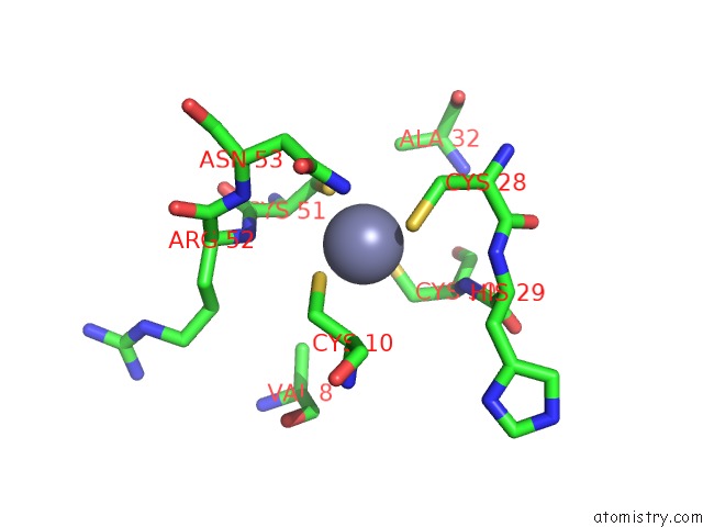
Zinc binding site 1 out of 4 in 4cl1
Go back to
Zinc binding site 1 out
of 4 in the The Crystal Structure of NS5A Domain 1 From Genotype 1A Reveals New Clues to the Mechanism of Action For Dimeric Hcv Inhibitors
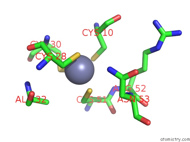
Mono view
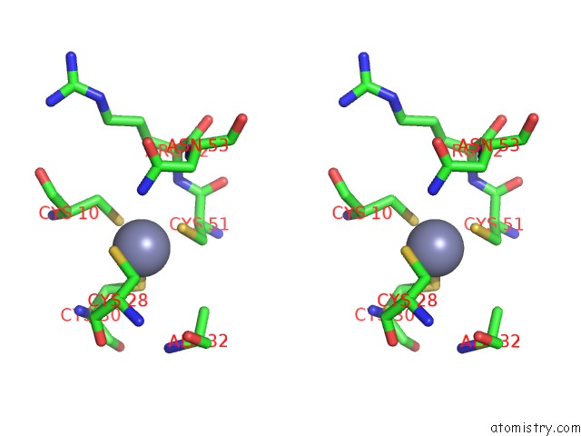
Stereo pair view
Mono view
Stereo pair view
A full contact list of Zinc with other atoms in the Zn binding
site number 1 of The Crystal Structure of NS5A Domain 1 From Genotype 1A Reveals New Clues to the Mechanism of Action For Dimeric Hcv Inhibitors within 5.0Å range:
|
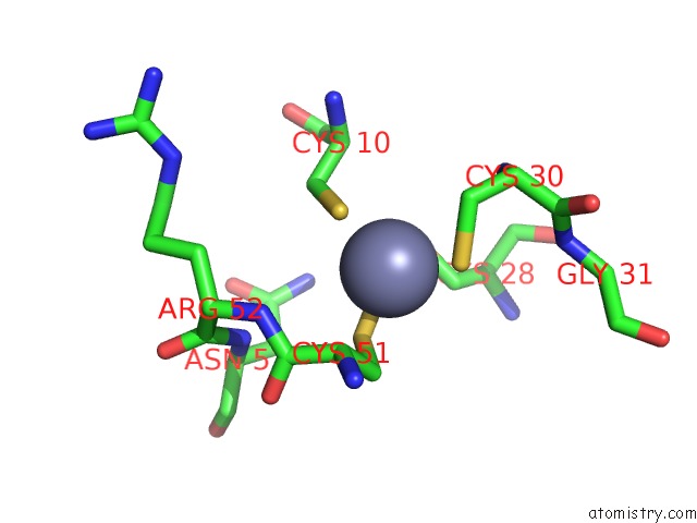
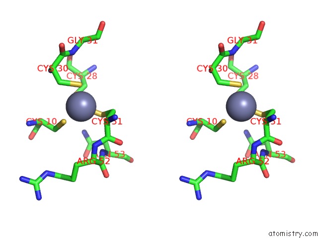
Zinc binding site 2 out of 4 in 4cl1
Go back to
Zinc binding site 2 out
of 4 in the The Crystal Structure of NS5A Domain 1 From Genotype 1A Reveals New Clues to the Mechanism of Action For Dimeric Hcv Inhibitors

Mono view

Stereo pair view
Mono view
Stereo pair view
A full contact list of Zinc with other atoms in the Zn binding
site number 2 of The Crystal Structure of NS5A Domain 1 From Genotype 1A Reveals New Clues to the Mechanism of Action For Dimeric Hcv Inhibitors within 5.0Å range:
|
Zinc binding site 3 out of 4 in 4cl1
Go back to
Zinc binding site 3 out
of 4 in the The Crystal Structure of NS5A Domain 1 From Genotype 1A Reveals New Clues to the Mechanism of Action For Dimeric Hcv Inhibitors
Mono view
Stereo pair view
Mono view
Stereo pair view
A full contact list of Zinc with other atoms in the Zn binding
site number 3 of The Crystal Structure of NS5A Domain 1 From Genotype 1A Reveals New Clues to the Mechanism of Action For Dimeric Hcv Inhibitors within 5.0Å range:
|
Zinc binding site 4 out of 4 in 4cl1
Go back to
Zinc binding site 4 out
of 4 in the The Crystal Structure of NS5A Domain 1 From Genotype 1A Reveals New Clues to the Mechanism of Action For Dimeric Hcv Inhibitors
Mono view

Stereo pair view
Mono view
Stereo pair view
A full contact list of Zinc with other atoms in the Zn binding
site number 4 of The Crystal Structure of NS5A Domain 1 From Genotype 1A Reveals New Clues to the Mechanism of Action For Dimeric Hcv Inhibitors within 5.0Å range:
|
Reference:
S.M.Lambert,
D.R.Langley,
J.A.Garnett,
R.Angell,
K.Hedgethorne,
N.A.Meanwell,
S.J.Matthews.
The Crystal Structure of NS5A Domain 1 From Genotype 1A Reveals New Clues to the Mechanism of Action For Dimeric Hcv Inhibitors. Protein Sci. V. 23 723 2014.
ISSN: ISSN 0961-8368
PubMed: 24639329
DOI: 10.1002/PRO.2456
Page generated: Sat Oct 26 20:48:38 2024
ISSN: ISSN 0961-8368
PubMed: 24639329
DOI: 10.1002/PRO.2456
Last articles
Zn in 9MJ5Zn in 9HNW
Zn in 9G0L
Zn in 9FNE
Zn in 9DZN
Zn in 9E0I
Zn in 9D32
Zn in 9DAK
Zn in 8ZXC
Zn in 8ZUF