Zinc »
PDB 4bjb-4bud »
4bt5 »
Zinc in PDB 4bt5: Acetolactate Decarboxylase with A Bound (2S,3R)-2,3- Dihydroxy-2-Methylbutanoic Acid
Enzymatic activity of Acetolactate Decarboxylase with A Bound (2S,3R)-2,3- Dihydroxy-2-Methylbutanoic Acid
All present enzymatic activity of Acetolactate Decarboxylase with A Bound (2S,3R)-2,3- Dihydroxy-2-Methylbutanoic Acid:
4.1.1.5;
4.1.1.5;
Protein crystallography data
The structure of Acetolactate Decarboxylase with A Bound (2S,3R)-2,3- Dihydroxy-2-Methylbutanoic Acid, PDB code: 4bt5
was solved by
V.A Marlow,
D.Rea,
S.Najmudin,
M.Wills,
V.Fulop,
with X-Ray Crystallography technique. A brief refinement statistics is given in the table below:
| Resolution Low / High (Å) | 34.74 / 1.10 |
| Space group | P 32 2 1 |
| Cell size a, b, c (Å), α, β, γ (°) | 47.130, 47.130, 198.630, 90.00, 90.00, 120.00 |
| R / Rfree (%) | 17.696 / 18.377 |
Zinc Binding Sites:
The binding sites of Zinc atom in the Acetolactate Decarboxylase with A Bound (2S,3R)-2,3- Dihydroxy-2-Methylbutanoic Acid
(pdb code 4bt5). This binding sites where shown within
5.0 Angstroms radius around Zinc atom.
In total only one binding site of Zinc was determined in the Acetolactate Decarboxylase with A Bound (2S,3R)-2,3- Dihydroxy-2-Methylbutanoic Acid, PDB code: 4bt5:
In total only one binding site of Zinc was determined in the Acetolactate Decarboxylase with A Bound (2S,3R)-2,3- Dihydroxy-2-Methylbutanoic Acid, PDB code: 4bt5:
Zinc binding site 1 out of 1 in 4bt5
Go back to
Zinc binding site 1 out
of 1 in the Acetolactate Decarboxylase with A Bound (2S,3R)-2,3- Dihydroxy-2-Methylbutanoic Acid
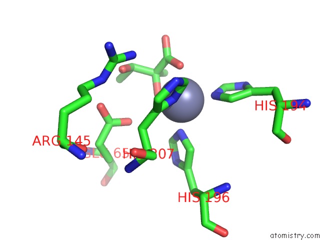
Mono view
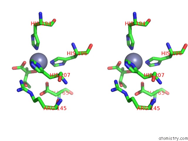
Stereo pair view
Mono view
Stereo pair view
A full contact list of Zinc with other atoms in the Zn binding
site number 1 of Acetolactate Decarboxylase with A Bound (2S,3R)-2,3- Dihydroxy-2-Methylbutanoic Acid within 5.0Å range:
|
Reference:
V.A.Marlow,
D.Rea,
S.Najmudin,
M.Wills,
V.Fulop.
Structure and Mechanism of Acetolactate Decarboxylase. Acs Chem.Biol. V. 8 2339 2013.
ISSN: ISSN 1554-8929
PubMed: 23985082
DOI: 10.1021/CB400429H
Page generated: Sat Oct 26 19:59:00 2024
ISSN: ISSN 1554-8929
PubMed: 23985082
DOI: 10.1021/CB400429H
Last articles
Zn in 9MJ5Zn in 9HNW
Zn in 9G0L
Zn in 9FNE
Zn in 9DZN
Zn in 9E0I
Zn in 9D32
Zn in 9DAK
Zn in 8ZXC
Zn in 8ZUF