Zinc »
PDB 4ad9-4arf »
4ai4 »
Zinc in PDB 4ai4: Crystal Structure of E38Q Mutant of 3-Methyladenine Dna Glycosylase I From Staphylococcus Aureus
Enzymatic activity of Crystal Structure of E38Q Mutant of 3-Methyladenine Dna Glycosylase I From Staphylococcus Aureus
All present enzymatic activity of Crystal Structure of E38Q Mutant of 3-Methyladenine Dna Glycosylase I From Staphylococcus Aureus:
3.2.2.20;
3.2.2.20;
Protein crystallography data
The structure of Crystal Structure of E38Q Mutant of 3-Methyladenine Dna Glycosylase I From Staphylococcus Aureus, PDB code: 4ai4
was solved by
X.Zhu,
J.H.Naismith,
with X-Ray Crystallography technique. A brief refinement statistics is given in the table below:
| Resolution Low / High (Å) | 53.67 / 1.73 |
| Space group | C 1 2 1 |
| Cell size a, b, c (Å), α, β, γ (°) | 107.060, 63.340, 38.230, 90.00, 109.26, 90.00 |
| R / Rfree (%) | 17.3 / 20.6 |
Zinc Binding Sites:
The binding sites of Zinc atom in the Crystal Structure of E38Q Mutant of 3-Methyladenine Dna Glycosylase I From Staphylococcus Aureus
(pdb code 4ai4). This binding sites where shown within
5.0 Angstroms radius around Zinc atom.
In total only one binding site of Zinc was determined in the Crystal Structure of E38Q Mutant of 3-Methyladenine Dna Glycosylase I From Staphylococcus Aureus, PDB code: 4ai4:
In total only one binding site of Zinc was determined in the Crystal Structure of E38Q Mutant of 3-Methyladenine Dna Glycosylase I From Staphylococcus Aureus, PDB code: 4ai4:
Zinc binding site 1 out of 1 in 4ai4
Go back to
Zinc binding site 1 out
of 1 in the Crystal Structure of E38Q Mutant of 3-Methyladenine Dna Glycosylase I From Staphylococcus Aureus
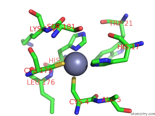
Mono view
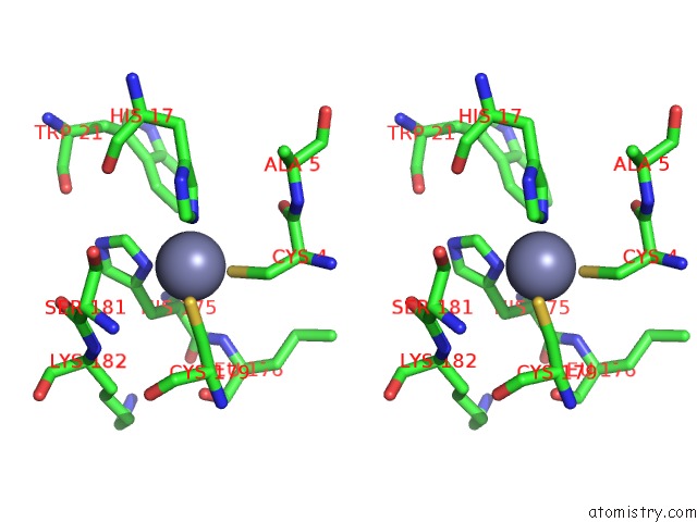
Stereo pair view
Mono view
Stereo pair view
A full contact list of Zinc with other atoms in the Zn binding
site number 1 of Crystal Structure of E38Q Mutant of 3-Methyladenine Dna Glycosylase I From Staphylococcus Aureus within 5.0Å range:
|
Reference:
X.Zhu,
X.Yan,
L.G.Carter,
H.Liu,
S.Graham,
P.J.Coote,
J.Naismith.
A Model For 3-Methyladenine Recognition By 3-Methyladenine Dna Glycosylase I (Tag) From Staphylococcus Aureus. Acta Crystallogr.,Sect.F V. 68 610 2012.
ISSN: ESSN 1744-3091
PubMed: 22684054
DOI: 10.1107/S1744309112016363
Page generated: Sat Oct 26 19:13:49 2024
ISSN: ESSN 1744-3091
PubMed: 22684054
DOI: 10.1107/S1744309112016363
Last articles
Zn in 9MJ5Zn in 9HNW
Zn in 9G0L
Zn in 9FNE
Zn in 9DZN
Zn in 9E0I
Zn in 9D32
Zn in 9DAK
Zn in 8ZXC
Zn in 8ZUF