Zinc »
PDB 3x17-3zr9 »
3zpc »
Zinc in PDB 3zpc: Acinetobacter Baumannii Ribd, Form 1
Enzymatic activity of Acinetobacter Baumannii Ribd, Form 1
Protein crystallography data
The structure of Acinetobacter Baumannii Ribd, Form 1, PDB code: 3zpc
was solved by
A.Dawson,
P.Trumper,
G.Chrysostomou,
W.N.Hunter,
with X-Ray Crystallography technique. A brief refinement statistics is given in the table below:
| Resolution Low / High (Å) | 117.14 / 2.20 |
| Space group | C 2 2 21 |
| Cell size a, b, c (Å), α, β, γ (°) | 148.987, 189.555, 76.406, 90.00, 90.00, 90.00 |
| R / Rfree (%) | 21.3 / 25 |
Zinc Binding Sites:
The binding sites of Zinc atom in the Acinetobacter Baumannii Ribd, Form 1
(pdb code 3zpc). This binding sites where shown within
5.0 Angstroms radius around Zinc atom.
In total 2 binding sites of Zinc where determined in the Acinetobacter Baumannii Ribd, Form 1, PDB code: 3zpc:
Jump to Zinc binding site number: 1; 2;
In total 2 binding sites of Zinc where determined in the Acinetobacter Baumannii Ribd, Form 1, PDB code: 3zpc:
Jump to Zinc binding site number: 1; 2;
Zinc binding site 1 out of 2 in 3zpc
Go back to
Zinc binding site 1 out
of 2 in the Acinetobacter Baumannii Ribd, Form 1
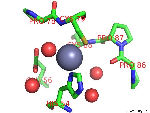
Mono view
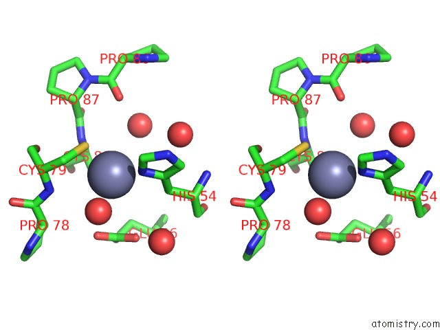
Stereo pair view
Mono view
Stereo pair view
A full contact list of Zinc with other atoms in the Zn binding
site number 1 of Acinetobacter Baumannii Ribd, Form 1 within 5.0Å range:
|
Zinc binding site 2 out of 2 in 3zpc
Go back to
Zinc binding site 2 out
of 2 in the Acinetobacter Baumannii Ribd, Form 1
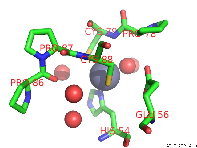
Mono view
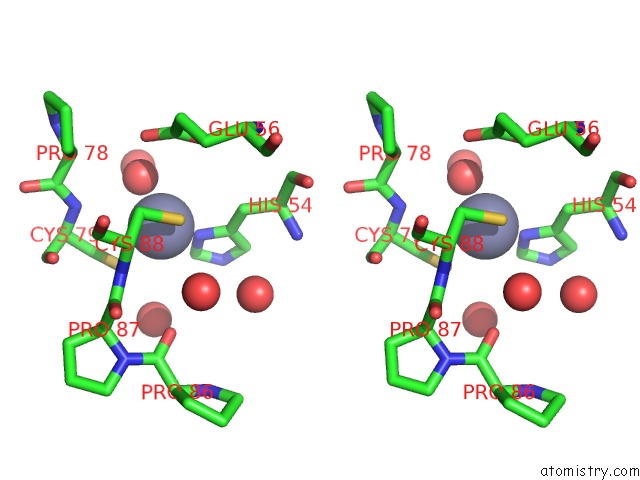
Stereo pair view
Mono view
Stereo pair view
A full contact list of Zinc with other atoms in the Zn binding
site number 2 of Acinetobacter Baumannii Ribd, Form 1 within 5.0Å range:
|
Reference:
A.Dawson,
P.Trumper,
G.Chrysostomou,
W.N.Hunter.
Structure of Diaminohydroxyphosphoribosylaminopyrimidine Deaminase/5-Amino-6-(5-Phosphoribosylamino)Uracil Reductase From Acinetobacter Baumannii. Acta Crystallogr.,Sect.F V. 69 611 2013.
ISSN: ESSN 1744-3091
PubMed: 23722836
DOI: 10.1107/S174430911301292X
Page generated: Sat Oct 26 18:31:42 2024
ISSN: ESSN 1744-3091
PubMed: 23722836
DOI: 10.1107/S174430911301292X
Last articles
Zn in 9MJ5Zn in 9HNW
Zn in 9G0L
Zn in 9FNE
Zn in 9DZN
Zn in 9E0I
Zn in 9D32
Zn in 9DAK
Zn in 8ZXC
Zn in 8ZUF