Zinc »
PDB 3vh4-3w5e »
3vyq »
Zinc in PDB 3vyq: Crystal Structure of the Methyl Cpg Binding Domain of MBD4 in Complex with the 5MCG/Tg Sequence in Space Group P1
Protein crystallography data
The structure of Crystal Structure of the Methyl Cpg Binding Domain of MBD4 in Complex with the 5MCG/Tg Sequence in Space Group P1, PDB code: 3vyq
was solved by
J.Otani,
K.Arita,
T.Kato,
M.Kinoshita,
M.Ariyoshi,
M.Shirakawa,
with X-Ray Crystallography technique. A brief refinement statistics is given in the table below:
| Resolution Low / High (Å) | 32.97 / 2.52 |
| Space group | P 1 |
| Cell size a, b, c (Å), α, β, γ (°) | 30.604, 34.093, 61.165, 75.28, 77.76, 87.31 |
| R / Rfree (%) | 19.3 / 23.6 |
Zinc Binding Sites:
The binding sites of Zinc atom in the Crystal Structure of the Methyl Cpg Binding Domain of MBD4 in Complex with the 5MCG/Tg Sequence in Space Group P1
(pdb code 3vyq). This binding sites where shown within
5.0 Angstroms radius around Zinc atom.
In total 3 binding sites of Zinc where determined in the Crystal Structure of the Methyl Cpg Binding Domain of MBD4 in Complex with the 5MCG/Tg Sequence in Space Group P1, PDB code: 3vyq:
Jump to Zinc binding site number: 1; 2; 3;
In total 3 binding sites of Zinc where determined in the Crystal Structure of the Methyl Cpg Binding Domain of MBD4 in Complex with the 5MCG/Tg Sequence in Space Group P1, PDB code: 3vyq:
Jump to Zinc binding site number: 1; 2; 3;
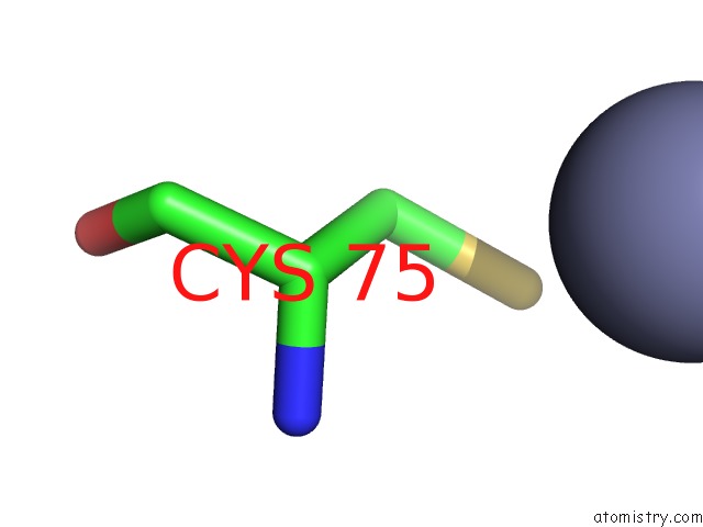
Zinc binding site 1 out of 3 in 3vyq
Go back to
Zinc binding site 1 out
of 3 in the Crystal Structure of the Methyl Cpg Binding Domain of MBD4 in Complex with the 5MCG/Tg Sequence in Space Group P1

Mono view

Stereo pair view
Mono view
Stereo pair view
A full contact list of Zinc with other atoms in the Zn binding
site number 1 of Crystal Structure of the Methyl Cpg Binding Domain of MBD4 in Complex with the 5MCG/Tg Sequence in Space Group P1 within 5.0Å range:
|
Zinc binding site 2 out of 3 in 3vyq
Go back to
Zinc binding site 2 out
of 3 in the Crystal Structure of the Methyl Cpg Binding Domain of MBD4 in Complex with the 5MCG/Tg Sequence in Space Group P1
Mono view

Stereo pair view
Mono view
Stereo pair view
A full contact list of Zinc with other atoms in the Zn binding
site number 2 of Crystal Structure of the Methyl Cpg Binding Domain of MBD4 in Complex with the 5MCG/Tg Sequence in Space Group P1 within 5.0Å range:
|
Zinc binding site 3 out of 3 in 3vyq
Go back to
Zinc binding site 3 out
of 3 in the Crystal Structure of the Methyl Cpg Binding Domain of MBD4 in Complex with the 5MCG/Tg Sequence in Space Group P1

Mono view

Stereo pair view
Mono view
Stereo pair view
A full contact list of Zinc with other atoms in the Zn binding
site number 3 of Crystal Structure of the Methyl Cpg Binding Domain of MBD4 in Complex with the 5MCG/Tg Sequence in Space Group P1 within 5.0Å range:
|
Reference:
J.Otani,
K.Arita,
T.Kato,
M.Kinoshita,
H.Kimura,
I.Suetake,
S.Tajima,
M.Ariyoshi,
M.Shirakawa.
Structural Basis of the Versatile Dna Recognition Ability of the Methyl-Cpg Binding Domain of Methyl-Cpg Binding Domain Protein 4 J.Biol.Chem. V. 288 6351 2013.
ISSN: ISSN 0021-9258
PubMed: 23316048
DOI: 10.1074/JBC.M112.431098
Page generated: Sat Oct 26 17:55:54 2024
ISSN: ISSN 0021-9258
PubMed: 23316048
DOI: 10.1074/JBC.M112.431098
Last articles
Zn in 9MJ5Zn in 9HNW
Zn in 9G0L
Zn in 9FNE
Zn in 9DZN
Zn in 9E0I
Zn in 9D32
Zn in 9DAK
Zn in 8ZXC
Zn in 8ZUF