Zinc »
PDB 3vh4-3w5e »
3vus »
Zinc in PDB 3vus: Escherichia Coli Pgab N-Terminal Domain
Protein crystallography data
The structure of Escherichia Coli Pgab N-Terminal Domain, PDB code: 3vus
was solved by
T.Nishiyama,
H.Noguchi,
H.Yoshida,
S.-Y.Park,
J.R.H.Tame,
with X-Ray Crystallography technique. A brief refinement statistics is given in the table below:
| Resolution Low / High (Å) | 25.00 / 1.65 |
| Space group | P 1 21 1 |
| Cell size a, b, c (Å), α, β, γ (°) | 39.559, 53.114, 144.170, 90.00, 95.25, 90.00 |
| R / Rfree (%) | 20.5 / 25.6 |
Other elements in 3vus:
The structure of Escherichia Coli Pgab N-Terminal Domain also contains other interesting chemical elements:
| Mercury | (Hg) | 1 atom |
Zinc Binding Sites:
The binding sites of Zinc atom in the Escherichia Coli Pgab N-Terminal Domain
(pdb code 3vus). This binding sites where shown within
5.0 Angstroms radius around Zinc atom.
In total 2 binding sites of Zinc where determined in the Escherichia Coli Pgab N-Terminal Domain, PDB code: 3vus:
Jump to Zinc binding site number: 1; 2;
In total 2 binding sites of Zinc where determined in the Escherichia Coli Pgab N-Terminal Domain, PDB code: 3vus:
Jump to Zinc binding site number: 1; 2;
Zinc binding site 1 out of 2 in 3vus
Go back to
Zinc binding site 1 out
of 2 in the Escherichia Coli Pgab N-Terminal Domain
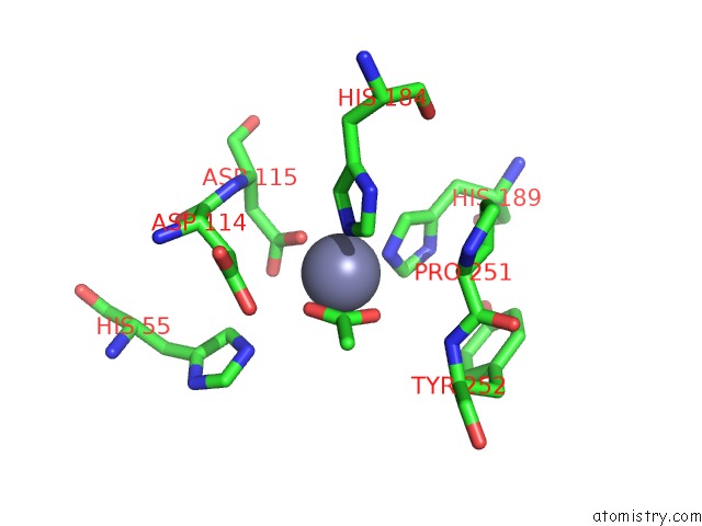
Mono view
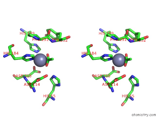
Stereo pair view
Mono view
Stereo pair view
A full contact list of Zinc with other atoms in the Zn binding
site number 1 of Escherichia Coli Pgab N-Terminal Domain within 5.0Å range:
|
Zinc binding site 2 out of 2 in 3vus
Go back to
Zinc binding site 2 out
of 2 in the Escherichia Coli Pgab N-Terminal Domain
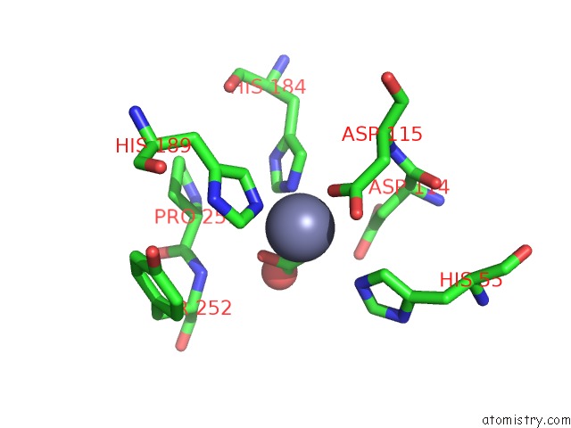
Mono view
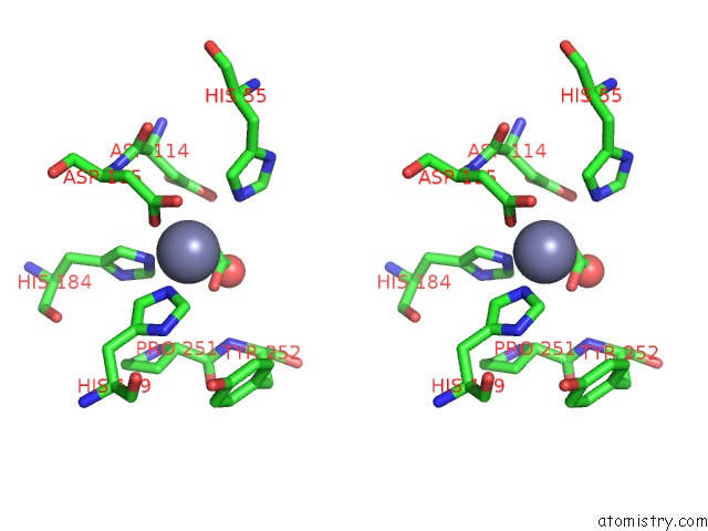
Stereo pair view
Mono view
Stereo pair view
A full contact list of Zinc with other atoms in the Zn binding
site number 2 of Escherichia Coli Pgab N-Terminal Domain within 5.0Å range:
|
Reference:
T.Nishiyama,
H.Noguchi,
H.Yoshida,
S.Y.Park,
J.R.Tame.
The Structure of the Deacetylase Domain of Escherichia Coli Pgab, An Enzyme Required For Biofilm Formation: A Circularly Permuted Member of the Carbohydrate Esterase 4 Family Acta Crystallogr.,Sect.D V. 69 44 2013.
ISSN: ISSN 0907-4449
PubMed: 23275162
DOI: 10.1107/S0907444912042059
Page generated: Sat Oct 26 17:53:24 2024
ISSN: ISSN 0907-4449
PubMed: 23275162
DOI: 10.1107/S0907444912042059
Last articles
Zn in 9MJ5Zn in 9HNW
Zn in 9G0L
Zn in 9FNE
Zn in 9DZN
Zn in 9E0I
Zn in 9D32
Zn in 9DAK
Zn in 8ZXC
Zn in 8ZUF