Zinc »
PDB 3tus-3u6p »
3tyo »
Zinc in PDB 3tyo: Structure of Neuronal Nitric Oxide Synthase Heme Domain in Complex with 6-(((3S,4S)-4-(2-((Furan-2-Ylmethyl)Amino)Ethoxy)Pyrrolidin-3- Yl)Methyl)-4-Methylpyridin-2-Amine
Enzymatic activity of Structure of Neuronal Nitric Oxide Synthase Heme Domain in Complex with 6-(((3S,4S)-4-(2-((Furan-2-Ylmethyl)Amino)Ethoxy)Pyrrolidin-3- Yl)Methyl)-4-Methylpyridin-2-Amine
All present enzymatic activity of Structure of Neuronal Nitric Oxide Synthase Heme Domain in Complex with 6-(((3S,4S)-4-(2-((Furan-2-Ylmethyl)Amino)Ethoxy)Pyrrolidin-3- Yl)Methyl)-4-Methylpyridin-2-Amine:
1.14.13.39;
1.14.13.39;
Protein crystallography data
The structure of Structure of Neuronal Nitric Oxide Synthase Heme Domain in Complex with 6-(((3S,4S)-4-(2-((Furan-2-Ylmethyl)Amino)Ethoxy)Pyrrolidin-3- Yl)Methyl)-4-Methylpyridin-2-Amine, PDB code: 3tyo
was solved by
H.Li,
T.L.Poulos,
with X-Ray Crystallography technique. A brief refinement statistics is given in the table below:
| Resolution Low / High (Å) | 37.90 / 1.93 |
| Space group | P 21 21 21 |
| Cell size a, b, c (Å), α, β, γ (°) | 51.934, 110.920, 164.316, 90.00, 90.00, 90.00 |
| R / Rfree (%) | 18 / 21.5 |
Other elements in 3tyo:
The structure of Structure of Neuronal Nitric Oxide Synthase Heme Domain in Complex with 6-(((3S,4S)-4-(2-((Furan-2-Ylmethyl)Amino)Ethoxy)Pyrrolidin-3- Yl)Methyl)-4-Methylpyridin-2-Amine also contains other interesting chemical elements:
| Iron | (Fe) | 2 atoms |
Zinc Binding Sites:
The binding sites of Zinc atom in the Structure of Neuronal Nitric Oxide Synthase Heme Domain in Complex with 6-(((3S,4S)-4-(2-((Furan-2-Ylmethyl)Amino)Ethoxy)Pyrrolidin-3- Yl)Methyl)-4-Methylpyridin-2-Amine
(pdb code 3tyo). This binding sites where shown within
5.0 Angstroms radius around Zinc atom.
In total only one binding site of Zinc was determined in the Structure of Neuronal Nitric Oxide Synthase Heme Domain in Complex with 6-(((3S,4S)-4-(2-((Furan-2-Ylmethyl)Amino)Ethoxy)Pyrrolidin-3- Yl)Methyl)-4-Methylpyridin-2-Amine, PDB code: 3tyo:
In total only one binding site of Zinc was determined in the Structure of Neuronal Nitric Oxide Synthase Heme Domain in Complex with 6-(((3S,4S)-4-(2-((Furan-2-Ylmethyl)Amino)Ethoxy)Pyrrolidin-3- Yl)Methyl)-4-Methylpyridin-2-Amine, PDB code: 3tyo:
Zinc binding site 1 out of 1 in 3tyo
Go back to
Zinc binding site 1 out
of 1 in the Structure of Neuronal Nitric Oxide Synthase Heme Domain in Complex with 6-(((3S,4S)-4-(2-((Furan-2-Ylmethyl)Amino)Ethoxy)Pyrrolidin-3- Yl)Methyl)-4-Methylpyridin-2-Amine
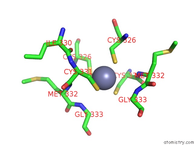
Mono view
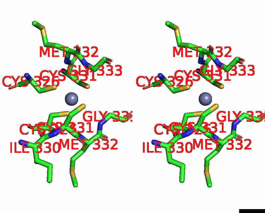
Stereo pair view
Mono view
Stereo pair view
A full contact list of Zinc with other atoms in the Zn binding
site number 1 of Structure of Neuronal Nitric Oxide Synthase Heme Domain in Complex with 6-(((3S,4S)-4-(2-((Furan-2-Ylmethyl)Amino)Ethoxy)Pyrrolidin-3- Yl)Methyl)-4-Methylpyridin-2-Amine within 5.0Å range:
|
Reference:
K.J.Labby,
F.Xue,
J.M.Kraus,
H.Ji,
J.Mataka,
H.Li,
P.Martasek,
L.J.Roman,
T.L.Poulos,
R.B.Silverman.
Intramolecular Hydrogen Bonding: A Potential Strategy For More Bioavailable Inhibitors of Neuronal Nitric Oxide Synthase. Bioorg.Med.Chem. V. 20 2435 2012.
ISSN: ISSN 0968-0896
PubMed: 22370337
DOI: 10.1016/J.BMC.2012.01.037
Page generated: Sat Oct 26 16:50:37 2024
ISSN: ISSN 0968-0896
PubMed: 22370337
DOI: 10.1016/J.BMC.2012.01.037
Last articles
Zn in 9MJ5Zn in 9HNW
Zn in 9G0L
Zn in 9FNE
Zn in 9DZN
Zn in 9E0I
Zn in 9D32
Zn in 9DAK
Zn in 8ZXC
Zn in 8ZUF