Zinc »
PDB 3tus-3u6p »
3two »
Zinc in PDB 3two: The Crystal Structure of Cad From Helicobacter Pylori Complexed with Nadp(H)
Enzymatic activity of The Crystal Structure of Cad From Helicobacter Pylori Complexed with Nadp(H)
All present enzymatic activity of The Crystal Structure of Cad From Helicobacter Pylori Complexed with Nadp(H):
1.1.1.195;
1.1.1.195;
Protein crystallography data
The structure of The Crystal Structure of Cad From Helicobacter Pylori Complexed with Nadp(H), PDB code: 3two
was solved by
K.H.Seo,
N.N.Zhuang,
C.Cong,
K.H.Lee,
with X-Ray Crystallography technique. A brief refinement statistics is given in the table below:
| Resolution Low / High (Å) | 43.31 / 2.18 |
| Space group | P 21 21 21 |
| Cell size a, b, c (Å), α, β, γ (°) | 85.410, 85.680, 100.491, 90.00, 90.00, 90.00 |
| R / Rfree (%) | 17.3 / 22.6 |
Zinc Binding Sites:
The binding sites of Zinc atom in the The Crystal Structure of Cad From Helicobacter Pylori Complexed with Nadp(H)
(pdb code 3two). This binding sites where shown within
5.0 Angstroms radius around Zinc atom.
In total 4 binding sites of Zinc where determined in the The Crystal Structure of Cad From Helicobacter Pylori Complexed with Nadp(H), PDB code: 3two:
Jump to Zinc binding site number: 1; 2; 3; 4;
In total 4 binding sites of Zinc where determined in the The Crystal Structure of Cad From Helicobacter Pylori Complexed with Nadp(H), PDB code: 3two:
Jump to Zinc binding site number: 1; 2; 3; 4;
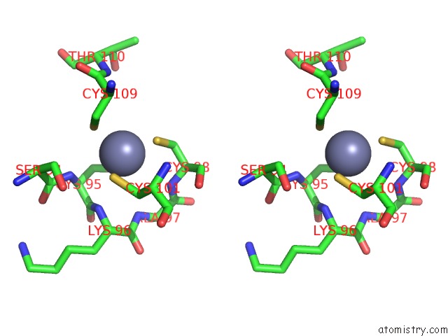
Zinc binding site 1 out of 4 in 3two
Go back to
Zinc binding site 1 out
of 4 in the The Crystal Structure of Cad From Helicobacter Pylori Complexed with Nadp(H)

Mono view

Stereo pair view
Mono view
Stereo pair view
A full contact list of Zinc with other atoms in the Zn binding
site number 1 of The Crystal Structure of Cad From Helicobacter Pylori Complexed with Nadp(H) within 5.0Å range:
|
Zinc binding site 2 out of 4 in 3two
Go back to
Zinc binding site 2 out
of 4 in the The Crystal Structure of Cad From Helicobacter Pylori Complexed with Nadp(H)

Mono view

Stereo pair view
Mono view
Stereo pair view
A full contact list of Zinc with other atoms in the Zn binding
site number 2 of The Crystal Structure of Cad From Helicobacter Pylori Complexed with Nadp(H) within 5.0Å range:
|
Zinc binding site 3 out of 4 in 3two
Go back to
Zinc binding site 3 out
of 4 in the The Crystal Structure of Cad From Helicobacter Pylori Complexed with Nadp(H)

Mono view

Stereo pair view
Mono view
Stereo pair view
A full contact list of Zinc with other atoms in the Zn binding
site number 3 of The Crystal Structure of Cad From Helicobacter Pylori Complexed with Nadp(H) within 5.0Å range:
|
Zinc binding site 4 out of 4 in 3two
Go back to
Zinc binding site 4 out
of 4 in the The Crystal Structure of Cad From Helicobacter Pylori Complexed with Nadp(H)

Mono view
Stereo pair view
Mono view
Stereo pair view
A full contact list of Zinc with other atoms in the Zn binding
site number 4 of The Crystal Structure of Cad From Helicobacter Pylori Complexed with Nadp(H) within 5.0Å range:
|
Reference:
K.H.Seo,
N.N.Zhuang,
C.Chen,
J.Y.Song,
H.L.Kang,
K.H.Rhee,
K.H.Lee.
Unusual Nadph Conformation in the Crystal Structure of A Cinnamyl Alcohol Dehydrogenase From Helicobacter Pylori in Complex with Nadp(H) and Substrate Docking Analysis Febs Lett. V. 586 337 2012.
ISSN: ISSN 0014-5793
PubMed: 22269576
DOI: 10.1016/J.FEBSLET.2012.01.020
Page generated: Sat Oct 26 16:49:02 2024
ISSN: ISSN 0014-5793
PubMed: 22269576
DOI: 10.1016/J.FEBSLET.2012.01.020
Last articles
Zn in 9MJ5Zn in 9HNW
Zn in 9G0L
Zn in 9FNE
Zn in 9DZN
Zn in 9E0I
Zn in 9D32
Zn in 9DAK
Zn in 8ZXC
Zn in 8ZUF