Zinc »
PDB 3tg5-3tty »
3ttf »
Zinc in PDB 3ttf: Crystal Structure of E. Coli Hypf with Amp and Carbamoyl Phosphate
Protein crystallography data
The structure of Crystal Structure of E. Coli Hypf with Amp and Carbamoyl Phosphate, PDB code: 3ttf
was solved by
S.Petkun,
R.Shi,
Y.Li,
M.Cygler,
with X-Ray Crystallography technique. A brief refinement statistics is given in the table below:
| Resolution Low / High (Å) | 100.33 / 1.92 |
| Space group | P 21 21 21 |
| Cell size a, b, c (Å), α, β, γ (°) | 46.361, 75.628, 200.668, 90.00, 90.00, 90.00 |
| R / Rfree (%) | 17.7 / 22.5 |
Other elements in 3ttf:
The structure of Crystal Structure of E. Coli Hypf with Amp and Carbamoyl Phosphate also contains other interesting chemical elements:
| Magnesium | (Mg) | 1 atom |
Zinc Binding Sites:
The binding sites of Zinc atom in the Crystal Structure of E. Coli Hypf with Amp and Carbamoyl Phosphate
(pdb code 3ttf). This binding sites where shown within
5.0 Angstroms radius around Zinc atom.
In total 3 binding sites of Zinc where determined in the Crystal Structure of E. Coli Hypf with Amp and Carbamoyl Phosphate, PDB code: 3ttf:
Jump to Zinc binding site number: 1; 2; 3;
In total 3 binding sites of Zinc where determined in the Crystal Structure of E. Coli Hypf with Amp and Carbamoyl Phosphate, PDB code: 3ttf:
Jump to Zinc binding site number: 1; 2; 3;
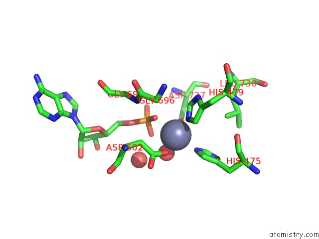
Zinc binding site 1 out of 3 in 3ttf
Go back to
Zinc binding site 1 out
of 3 in the Crystal Structure of E. Coli Hypf with Amp and Carbamoyl Phosphate
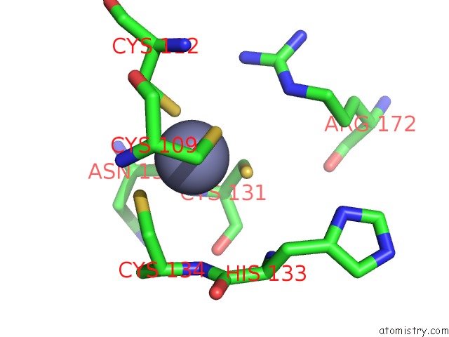
Mono view
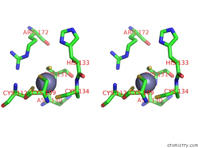
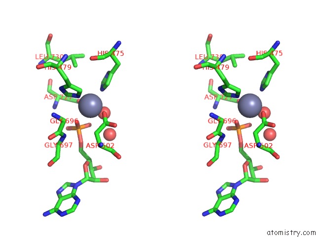
Stereo pair view
Mono view
Stereo pair view
A full contact list of Zinc with other atoms in the Zn binding
site number 1 of Crystal Structure of E. Coli Hypf with Amp and Carbamoyl Phosphate within 5.0Å range:
|
Zinc binding site 2 out of 3 in 3ttf
Go back to
Zinc binding site 2 out
of 3 in the Crystal Structure of E. Coli Hypf with Amp and Carbamoyl Phosphate
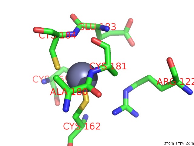
Mono view
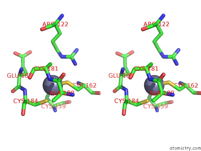
Stereo pair view
Mono view
Stereo pair view
A full contact list of Zinc with other atoms in the Zn binding
site number 2 of Crystal Structure of E. Coli Hypf with Amp and Carbamoyl Phosphate within 5.0Å range:
|
Zinc binding site 3 out of 3 in 3ttf
Go back to
Zinc binding site 3 out
of 3 in the Crystal Structure of E. Coli Hypf with Amp and Carbamoyl Phosphate
Mono view
Stereo pair view
Mono view
Stereo pair view
A full contact list of Zinc with other atoms in the Zn binding
site number 3 of Crystal Structure of E. Coli Hypf with Amp and Carbamoyl Phosphate within 5.0Å range:
|
Reference:
S.Petkun,
R.Shi,
Y.Li,
A.Asinas,
C.Munger,
L.Zhang,
M.Waclawek,
B.Soboh,
R.G.Sawers,
M.Cygler.
Structure of Hydrogenase Maturation Protein Hypf with Reaction Intermediates Shows Two Active Sites. Structure V. 19 1773 2011.
ISSN: ISSN 0969-2126
PubMed: 22153500
DOI: 10.1016/J.STR.2011.09.023
Page generated: Sat Oct 26 16:45:51 2024
ISSN: ISSN 0969-2126
PubMed: 22153500
DOI: 10.1016/J.STR.2011.09.023
Last articles
Zn in 9MJ5Zn in 9HNW
Zn in 9G0L
Zn in 9FNE
Zn in 9DZN
Zn in 9E0I
Zn in 9D32
Zn in 9DAK
Zn in 8ZXC
Zn in 8ZUF