Zinc »
PDB 3tg5-3tty »
3tmn »
Zinc in PDB 3tmn: The Binding of L-Valyl-L-Tryptophan to Crystalline Thermolysin Illustrates the Mode of Interaction of A Product of Peptide Hydrolysis
Enzymatic activity of The Binding of L-Valyl-L-Tryptophan to Crystalline Thermolysin Illustrates the Mode of Interaction of A Product of Peptide Hydrolysis
All present enzymatic activity of The Binding of L-Valyl-L-Tryptophan to Crystalline Thermolysin Illustrates the Mode of Interaction of A Product of Peptide Hydrolysis:
3.4.24.27;
3.4.24.27;
Protein crystallography data
The structure of The Binding of L-Valyl-L-Tryptophan to Crystalline Thermolysin Illustrates the Mode of Interaction of A Product of Peptide Hydrolysis, PDB code: 3tmn
was solved by
H.M.Holden,
B.W.Matthews,
with X-Ray Crystallography technique. A brief refinement statistics is given in the table below:
| Resolution Low / High (Å) | 10.00 / 1.70 |
| Space group | P 61 2 2 |
| Cell size a, b, c (Å), α, β, γ (°) | 94.100, 94.100, 131.400, 90.00, 90.00, 120.00 |
| R / Rfree (%) | n/a / n/a |
Other elements in 3tmn:
The structure of The Binding of L-Valyl-L-Tryptophan to Crystalline Thermolysin Illustrates the Mode of Interaction of A Product of Peptide Hydrolysis also contains other interesting chemical elements:
| Calcium | (Ca) | 4 atoms |
Zinc Binding Sites:
The binding sites of Zinc atom in the The Binding of L-Valyl-L-Tryptophan to Crystalline Thermolysin Illustrates the Mode of Interaction of A Product of Peptide Hydrolysis
(pdb code 3tmn). This binding sites where shown within
5.0 Angstroms radius around Zinc atom.
In total only one binding site of Zinc was determined in the The Binding of L-Valyl-L-Tryptophan to Crystalline Thermolysin Illustrates the Mode of Interaction of A Product of Peptide Hydrolysis, PDB code: 3tmn:
In total only one binding site of Zinc was determined in the The Binding of L-Valyl-L-Tryptophan to Crystalline Thermolysin Illustrates the Mode of Interaction of A Product of Peptide Hydrolysis, PDB code: 3tmn:
Zinc binding site 1 out of 1 in 3tmn
Go back to
Zinc binding site 1 out
of 1 in the The Binding of L-Valyl-L-Tryptophan to Crystalline Thermolysin Illustrates the Mode of Interaction of A Product of Peptide Hydrolysis
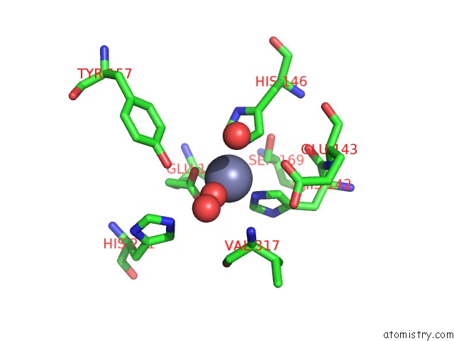
Mono view
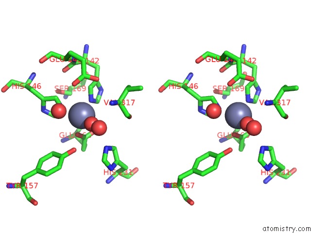
Stereo pair view
Mono view
Stereo pair view
A full contact list of Zinc with other atoms in the Zn binding
site number 1 of The Binding of L-Valyl-L-Tryptophan to Crystalline Thermolysin Illustrates the Mode of Interaction of A Product of Peptide Hydrolysis within 5.0Å range:
|
Reference:
H.M.Holden,
B.W.Matthews.
The Binding of L-Valyl-L-Tryptophan to Crystalline Thermolysin Illustrates the Mode of Interaction of A Product of Peptide Hydrolysis. J.Biol.Chem. V. 263 3256 1988.
ISSN: ISSN 0021-9258
PubMed: 3343246
Page generated: Sat Oct 26 16:38:42 2024
ISSN: ISSN 0021-9258
PubMed: 3343246
Last articles
Zn in 9MJ5Zn in 9HNW
Zn in 9G0L
Zn in 9FNE
Zn in 9DZN
Zn in 9E0I
Zn in 9D32
Zn in 9DAK
Zn in 8ZXC
Zn in 8ZUF