Zinc »
PDB 3phx-3ptm »
3pki »
Zinc in PDB 3pki: Human SIRT6 Crystal Structure in Complex with Adp Ribose
Protein crystallography data
The structure of Human SIRT6 Crystal Structure in Complex with Adp Ribose, PDB code: 3pki
was solved by
P.W.Pan,
A.Dong,
W.Qiu,
P.Loppnau,
J.Wang,
M.Ravichandran,
A.Bochkarev,
C.Bountra,
J.Weigelt,
C.H.Arrowsmith,
J.Min,
A.M.Edwards,
Structuralgenomics Consortium (Sgc),
with X-Ray Crystallography technique. A brief refinement statistics is given in the table below:
| Resolution Low / High (Å) | 29.87 / 2.04 |
| Space group | P 1 |
| Cell size a, b, c (Å), α, β, γ (°) | 77.367, 90.200, 90.180, 118.09, 91.39, 115.80 |
| R / Rfree (%) | 18.3 / 21.3 |
Zinc Binding Sites:
The binding sites of Zinc atom in the Human SIRT6 Crystal Structure in Complex with Adp Ribose
(pdb code 3pki). This binding sites where shown within
5.0 Angstroms radius around Zinc atom.
In total 6 binding sites of Zinc where determined in the Human SIRT6 Crystal Structure in Complex with Adp Ribose, PDB code: 3pki:
Jump to Zinc binding site number: 1; 2; 3; 4; 5; 6;
In total 6 binding sites of Zinc where determined in the Human SIRT6 Crystal Structure in Complex with Adp Ribose, PDB code: 3pki:
Jump to Zinc binding site number: 1; 2; 3; 4; 5; 6;
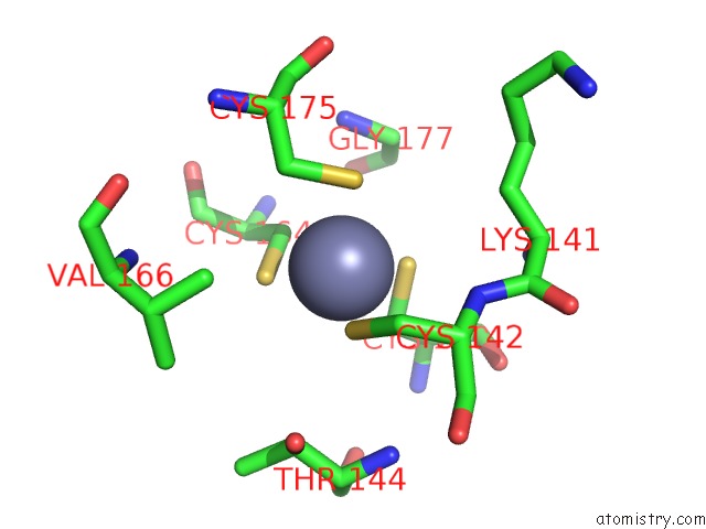
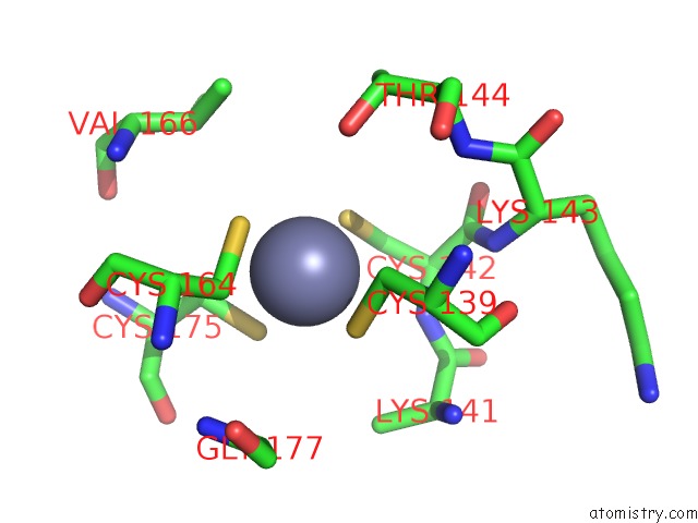
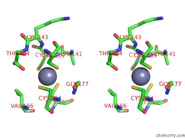
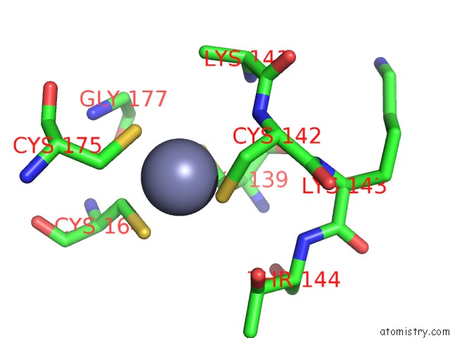
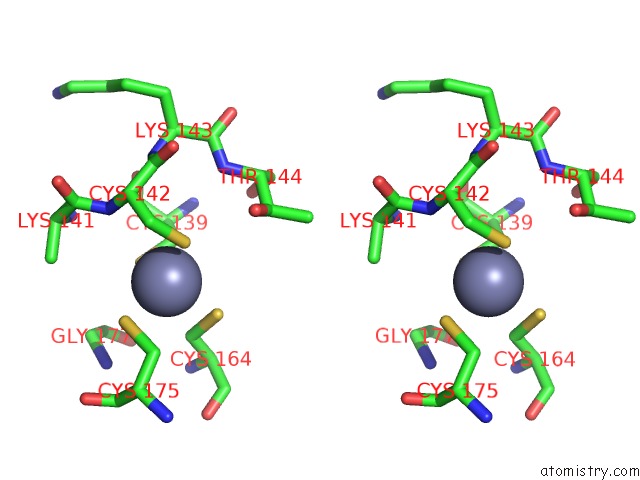
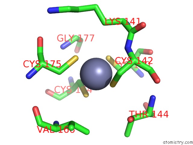
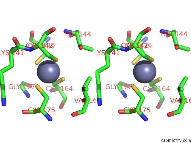
Zinc binding site 1 out of 6 in 3pki
Go back to
Zinc binding site 1 out
of 6 in the Human SIRT6 Crystal Structure in Complex with Adp Ribose
Mono view
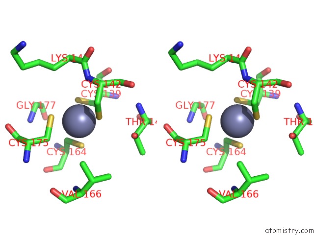
Stereo pair view
Mono view
Stereo pair view
A full contact list of Zinc with other atoms in the Zn binding
site number 1 of Human SIRT6 Crystal Structure in Complex with Adp Ribose within 5.0Å range:
|
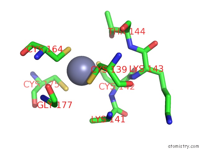
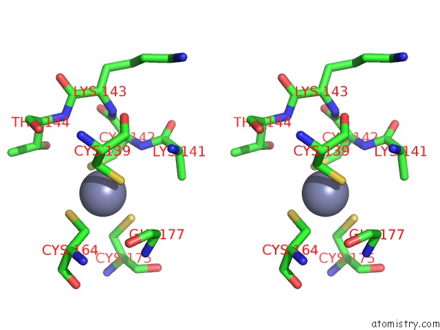
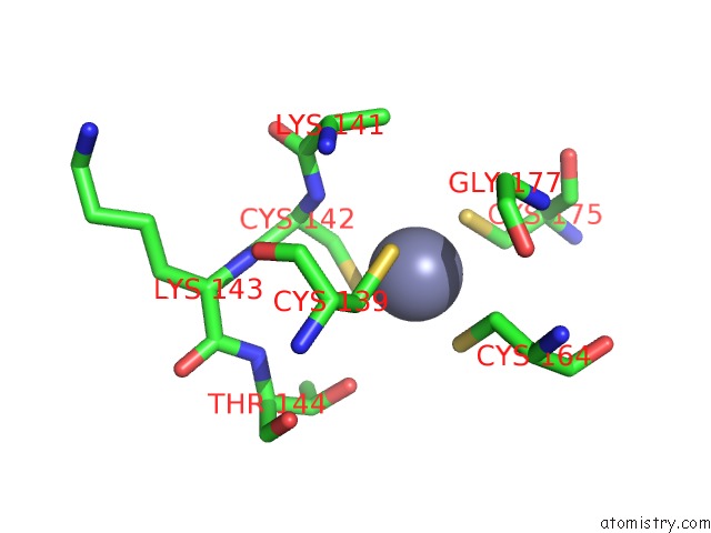
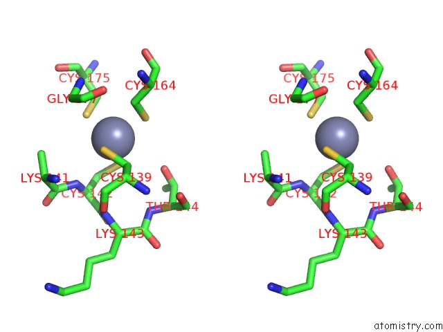
Zinc binding site 2 out of 6 in 3pki
Go back to
Zinc binding site 2 out
of 6 in the Human SIRT6 Crystal Structure in Complex with Adp Ribose
Mono view
Stereo pair view
Mono view
Stereo pair view
A full contact list of Zinc with other atoms in the Zn binding
site number 2 of Human SIRT6 Crystal Structure in Complex with Adp Ribose within 5.0Å range:
|
Zinc binding site 3 out of 6 in 3pki
Go back to
Zinc binding site 3 out
of 6 in the Human SIRT6 Crystal Structure in Complex with Adp Ribose
Mono view
Stereo pair view
Mono view
Stereo pair view
A full contact list of Zinc with other atoms in the Zn binding
site number 3 of Human SIRT6 Crystal Structure in Complex with Adp Ribose within 5.0Å range:
|
Zinc binding site 4 out of 6 in 3pki
Go back to
Zinc binding site 4 out
of 6 in the Human SIRT6 Crystal Structure in Complex with Adp Ribose
Mono view
Stereo pair view
Mono view
Stereo pair view
A full contact list of Zinc with other atoms in the Zn binding
site number 4 of Human SIRT6 Crystal Structure in Complex with Adp Ribose within 5.0Å range:
|
Zinc binding site 5 out of 6 in 3pki
Go back to
Zinc binding site 5 out
of 6 in the Human SIRT6 Crystal Structure in Complex with Adp Ribose
Mono view
Stereo pair view
Mono view
Stereo pair view
A full contact list of Zinc with other atoms in the Zn binding
site number 5 of Human SIRT6 Crystal Structure in Complex with Adp Ribose within 5.0Å range:
|
Zinc binding site 6 out of 6 in 3pki
Go back to
Zinc binding site 6 out
of 6 in the Human SIRT6 Crystal Structure in Complex with Adp Ribose
Mono view
Stereo pair view
Mono view
Stereo pair view
A full contact list of Zinc with other atoms in the Zn binding
site number 6 of Human SIRT6 Crystal Structure in Complex with Adp Ribose within 5.0Å range:
|
Reference:
P.W.Pan,
J.L.Feldman,
M.K.Devries,
A.Dong,
A.M.Edwards,
J.M.Denu.
Structure and Biochemical Functions of SIRT6. J.Biol.Chem. V. 286 14575 2011.
ISSN: ISSN 0021-9258
PubMed: 21362626
DOI: 10.1074/JBC.M111.218990
Page generated: Sat Oct 26 11:27:03 2024
ISSN: ISSN 0021-9258
PubMed: 21362626
DOI: 10.1074/JBC.M111.218990
Last articles
Zn in 9MJ5Zn in 9HNW
Zn in 9G0L
Zn in 9FNE
Zn in 9DZN
Zn in 9E0I
Zn in 9D32
Zn in 9DAK
Zn in 8ZXC
Zn in 8ZUF