Zinc »
PDB 3phx-3ptm »
3pjj »
Zinc in PDB 3pjj: Synthetic Dimer of Human Carbonic Anhydrase II
Enzymatic activity of Synthetic Dimer of Human Carbonic Anhydrase II
All present enzymatic activity of Synthetic Dimer of Human Carbonic Anhydrase II:
4.2.1.1;
4.2.1.1;
Protein crystallography data
The structure of Synthetic Dimer of Human Carbonic Anhydrase II, PDB code: 3pjj
was solved by
P.W.Snyder,
R.L.Kwant,
D.T.Moustakas,
E.T.Mack,
M.J.Butte,
G.W.Whitesides,
with X-Ray Crystallography technique. A brief refinement statistics is given in the table below:
| Resolution Low / High (Å) | 50.00 / 1.80 |
| Space group | C 1 2 1 |
| Cell size a, b, c (Å), α, β, γ (°) | 66.705, 51.132, 80.935, 90.00, 107.19, 90.00 |
| R / Rfree (%) | 21.9 / 28.3 |
Zinc Binding Sites:
The binding sites of Zinc atom in the Synthetic Dimer of Human Carbonic Anhydrase II
(pdb code 3pjj). This binding sites where shown within
5.0 Angstroms radius around Zinc atom.
In total only one binding site of Zinc was determined in the Synthetic Dimer of Human Carbonic Anhydrase II, PDB code: 3pjj:
In total only one binding site of Zinc was determined in the Synthetic Dimer of Human Carbonic Anhydrase II, PDB code: 3pjj:
Zinc binding site 1 out of 1 in 3pjj
Go back to
Zinc binding site 1 out
of 1 in the Synthetic Dimer of Human Carbonic Anhydrase II

Mono view
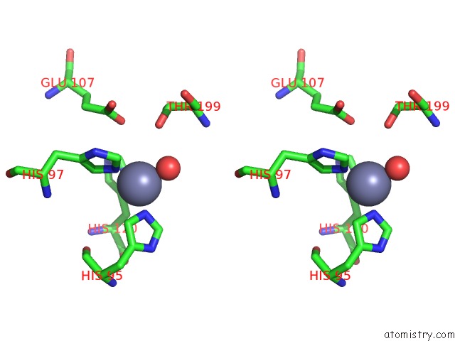
Stereo pair view
Mono view
Stereo pair view
A full contact list of Zinc with other atoms in the Zn binding
site number 1 of Synthetic Dimer of Human Carbonic Anhydrase II within 5.0Å range:
|
Reference:
P.W.Snyder,
R.L.Kwant,
D.T.Moustakas,
E.T.Mack,
M.J.Butte,
G.W.Whitesides.
One Interface Stabilizes Linear Chains in All Polymorphs of Crystals of Human Carbonic Anhydrase II To Be Published.
Page generated: Sat Oct 26 11:27:02 2024
Last articles
Zn in 9MJ5Zn in 9HNW
Zn in 9G0L
Zn in 9FNE
Zn in 9DZN
Zn in 9E0I
Zn in 9D32
Zn in 9DAK
Zn in 8ZXC
Zn in 8ZUF