Zinc »
PDB 3n3k-3n9o »
3n6w »
Zinc in PDB 3n6w: Crystal Structure of Human Gamma-Butyrobetaine Hydroxylase
Enzymatic activity of Crystal Structure of Human Gamma-Butyrobetaine Hydroxylase
All present enzymatic activity of Crystal Structure of Human Gamma-Butyrobetaine Hydroxylase:
1.14.11.1;
1.14.11.1;
Protein crystallography data
The structure of Crystal Structure of Human Gamma-Butyrobetaine Hydroxylase, PDB code: 3n6w
was solved by
J.Rumnieks,
A.Zeltins,
A.Leonchiks,
A.Kazaks,
S.Kotelovica,
K.Tars,
with X-Ray Crystallography technique. A brief refinement statistics is given in the table below:
| Resolution Low / High (Å) | 142.72 / 2.00 |
| Space group | P 65 2 2 |
| Cell size a, b, c (Å), α, β, γ (°) | 164.800, 164.800, 97.780, 90.00, 90.00, 120.00 |
| R / Rfree (%) | 20.8 / 23.3 |
Zinc Binding Sites:
The binding sites of Zinc atom in the Crystal Structure of Human Gamma-Butyrobetaine Hydroxylase
(pdb code 3n6w). This binding sites where shown within
5.0 Angstroms radius around Zinc atom.
In total only one binding site of Zinc was determined in the Crystal Structure of Human Gamma-Butyrobetaine Hydroxylase, PDB code: 3n6w:
In total only one binding site of Zinc was determined in the Crystal Structure of Human Gamma-Butyrobetaine Hydroxylase, PDB code: 3n6w:
Zinc binding site 1 out of 1 in 3n6w
Go back to
Zinc binding site 1 out
of 1 in the Crystal Structure of Human Gamma-Butyrobetaine Hydroxylase
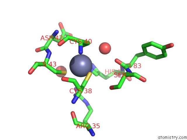
Mono view
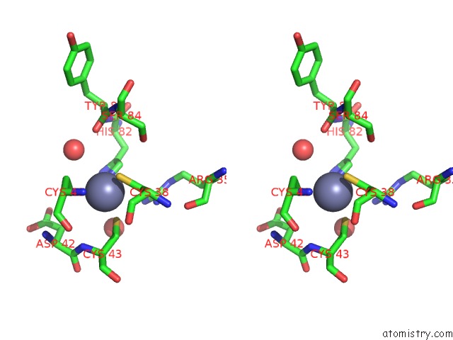
Stereo pair view
Mono view
Stereo pair view
A full contact list of Zinc with other atoms in the Zn binding
site number 1 of Crystal Structure of Human Gamma-Butyrobetaine Hydroxylase within 5.0Å range:
|
Reference:
K.Tars,
J.Rumnieks,
A.Zeltins,
A.Kazaks,
S.Kotelovica,
A.Leonciks,
J.Sharipo,
A.Viksna,
J.Kuka,
E.Liepinsh,
M.Dambrova.
Crystal Structure of Human Gamma-Butyrobetaine Hydroxylase. Biochem.Biophys.Res.Commun. V. 398 634 2010.
ISSN: ISSN 0006-291X
PubMed: 20599753
DOI: 10.1016/J.BBRC.2010.06.121
Page generated: Sat Oct 26 10:06:27 2024
ISSN: ISSN 0006-291X
PubMed: 20599753
DOI: 10.1016/J.BBRC.2010.06.121
Last articles
Zn in 9MJ5Zn in 9HNW
Zn in 9G0L
Zn in 9FNE
Zn in 9DZN
Zn in 9E0I
Zn in 9D32
Zn in 9DAK
Zn in 8ZXC
Zn in 8ZUF