Zinc »
PDB 3hk5-3hqy »
3hk7 »
Zinc in PDB 3hk7: Crystal Structure of Uronate Isomerase From Bacillus Halodurans Complexed with Zinc and D-Arabinarate, Monoclinic Crystal Form
Protein crystallography data
The structure of Crystal Structure of Uronate Isomerase From Bacillus Halodurans Complexed with Zinc and D-Arabinarate, Monoclinic Crystal Form, PDB code: 3hk7
was solved by
A.A.Fedorov,
E.V.Fedorov,
T.T.Nguyen,
F.M.Raushel,
S.C.Almo,
with X-Ray Crystallography technique. A brief refinement statistics is given in the table below:
| Resolution Low / High (Å) | 24.99 / 2.20 |
| Space group | C 1 2 1 |
| Cell size a, b, c (Å), α, β, γ (°) | 274.823, 156.516, 185.959, 90.00, 116.20, 90.00 |
| R / Rfree (%) | 21.3 / 24.5 |
Other elements in 3hk7:
The structure of Crystal Structure of Uronate Isomerase From Bacillus Halodurans Complexed with Zinc and D-Arabinarate, Monoclinic Crystal Form also contains other interesting chemical elements:
| Chlorine | (Cl) | 4 atoms |
| Sodium | (Na) | 4 atoms |
Zinc Binding Sites:
Pages:
>>> Page 1 <<< Page 2, Binding sites: 11 - 12;Binding sites:
The binding sites of Zinc atom in the Crystal Structure of Uronate Isomerase From Bacillus Halodurans Complexed with Zinc and D-Arabinarate, Monoclinic Crystal Form (pdb code 3hk7). This binding sites where shown within 5.0 Angstroms radius around Zinc atom.In total 12 binding sites of Zinc where determined in the Crystal Structure of Uronate Isomerase From Bacillus Halodurans Complexed with Zinc and D-Arabinarate, Monoclinic Crystal Form, PDB code: 3hk7:
Jump to Zinc binding site number: 1; 2; 3; 4; 5; 6; 7; 8; 9; 10;
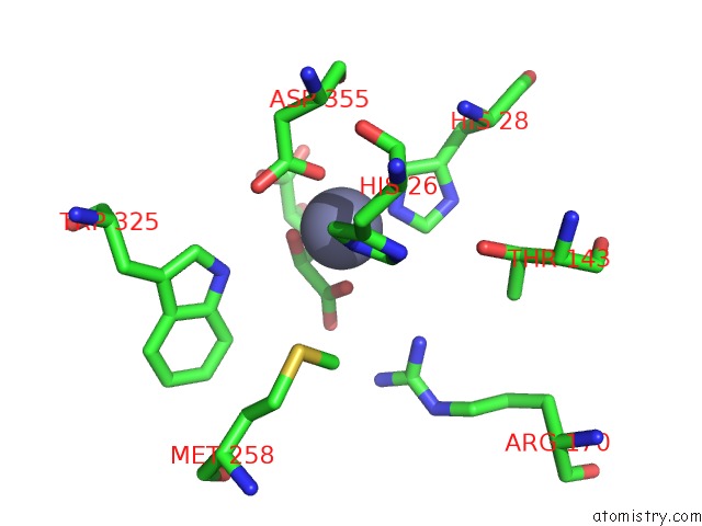
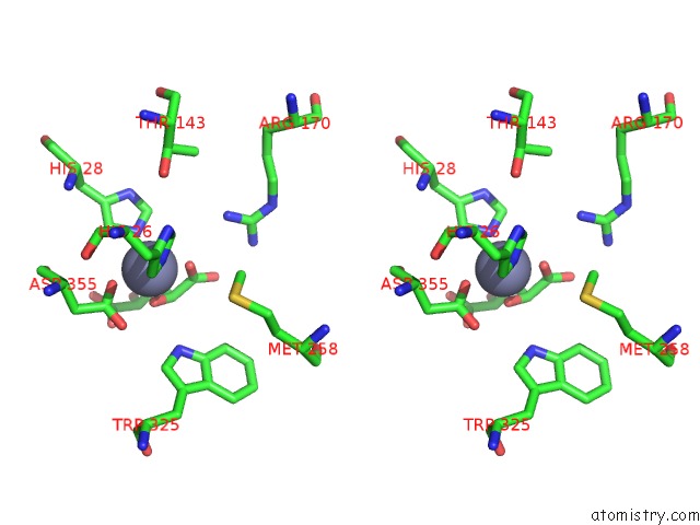
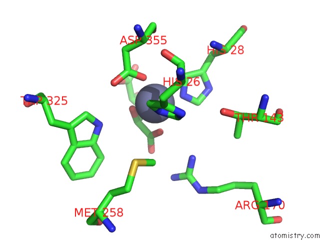
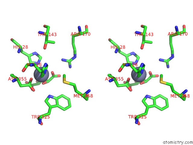
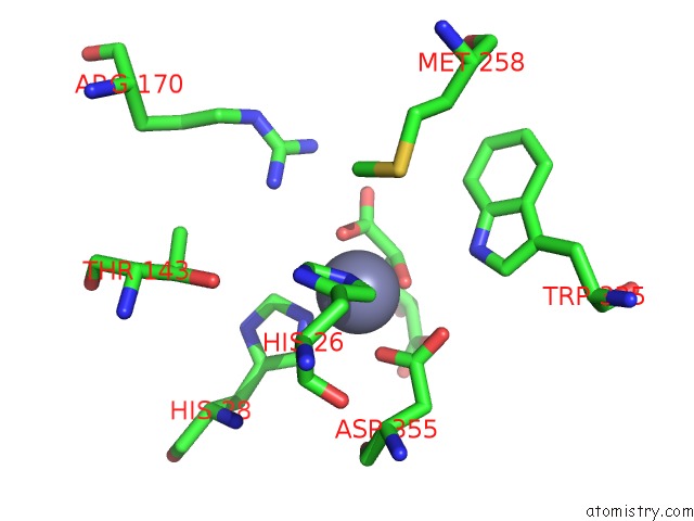
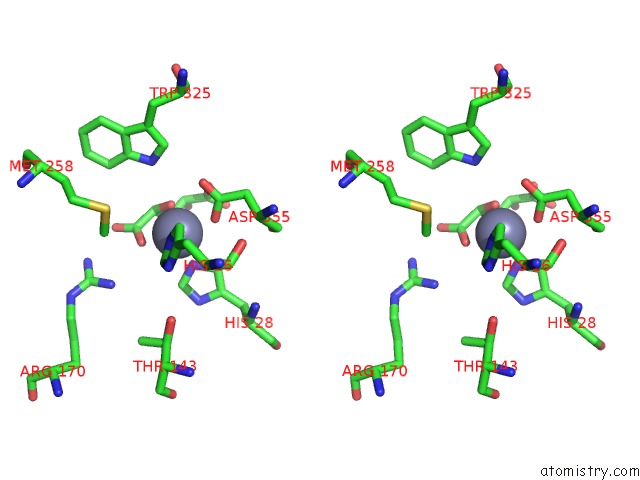
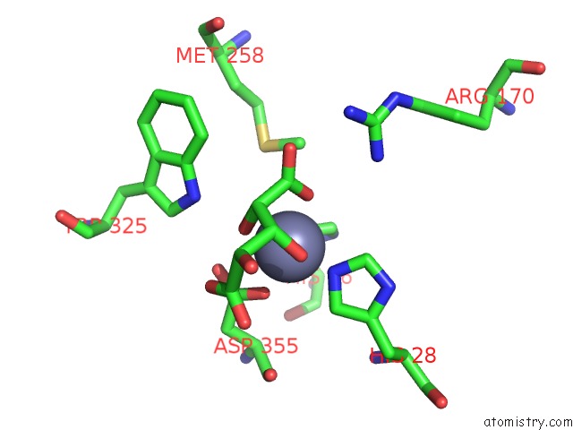
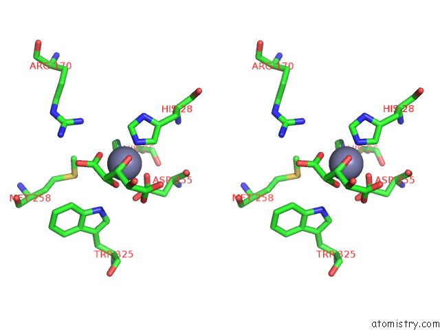
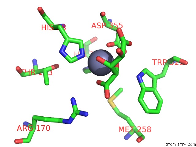
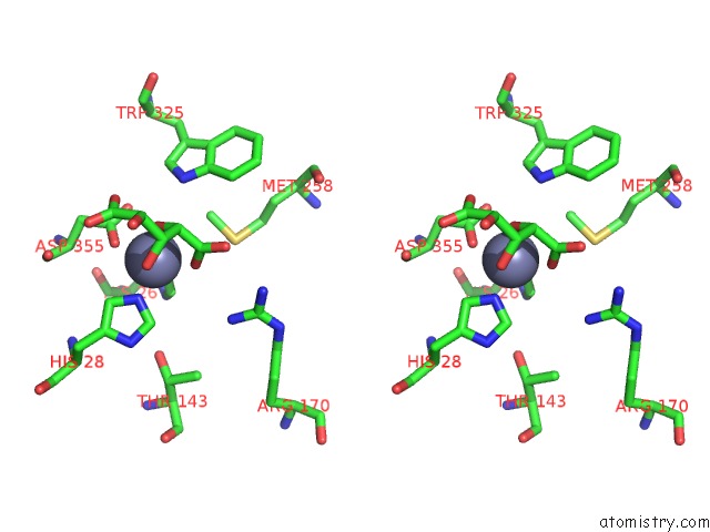
Zinc binding site 1 out of 12 in 3hk7
Go back to
Zinc binding site 1 out
of 12 in the Crystal Structure of Uronate Isomerase From Bacillus Halodurans Complexed with Zinc and D-Arabinarate, Monoclinic Crystal Form
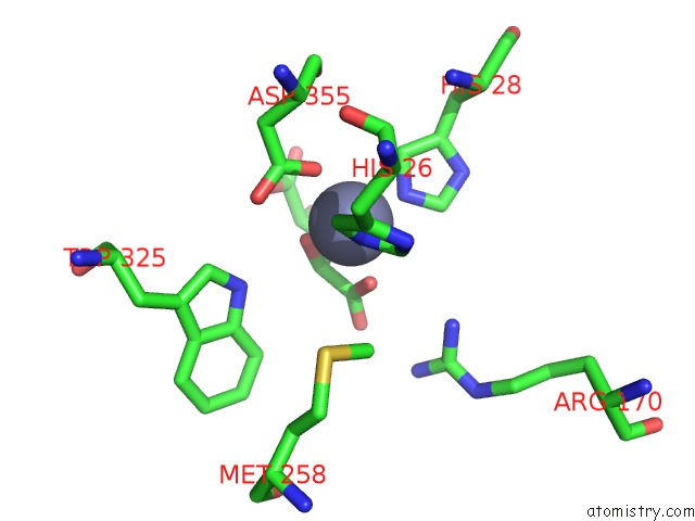
Mono view
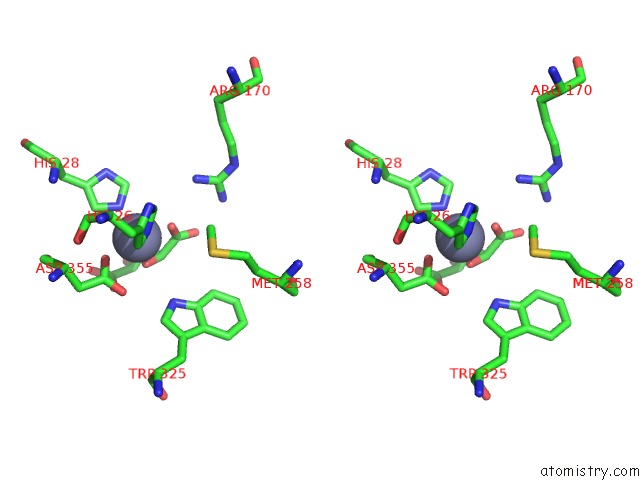
Stereo pair view
Mono view
Stereo pair view
A full contact list of Zinc with other atoms in the Zn binding
site number 1 of Crystal Structure of Uronate Isomerase From Bacillus Halodurans Complexed with Zinc and D-Arabinarate, Monoclinic Crystal Form within 5.0Å range:
|
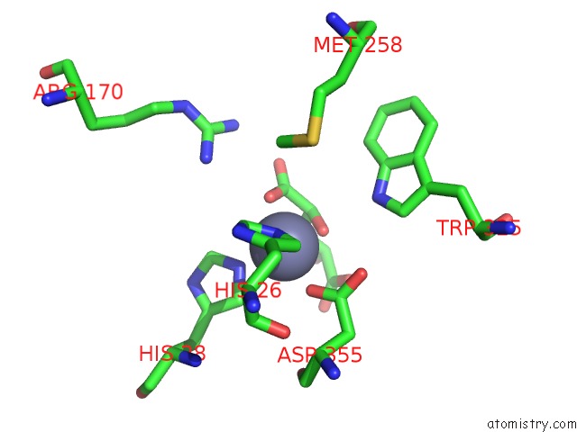
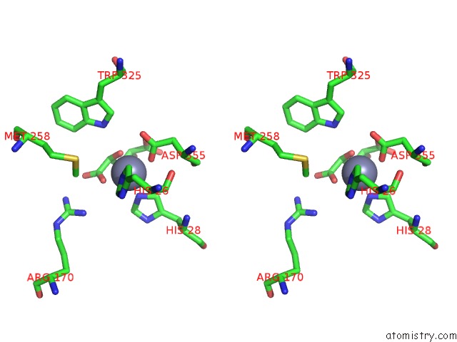
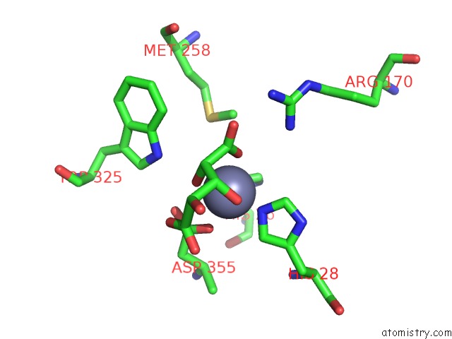
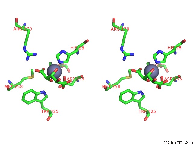
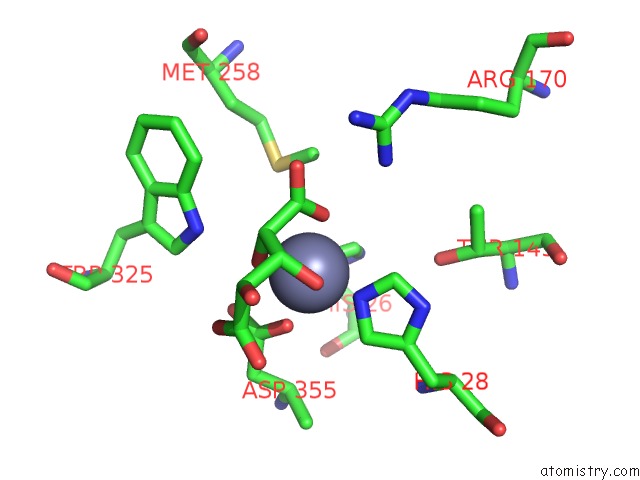
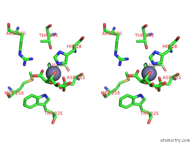
Zinc binding site 2 out of 12 in 3hk7
Go back to
Zinc binding site 2 out
of 12 in the Crystal Structure of Uronate Isomerase From Bacillus Halodurans Complexed with Zinc and D-Arabinarate, Monoclinic Crystal Form
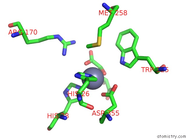
Mono view
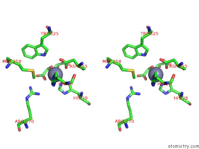
Stereo pair view
Mono view
Stereo pair view
A full contact list of Zinc with other atoms in the Zn binding
site number 2 of Crystal Structure of Uronate Isomerase From Bacillus Halodurans Complexed with Zinc and D-Arabinarate, Monoclinic Crystal Form within 5.0Å range:
|
Zinc binding site 3 out of 12 in 3hk7
Go back to
Zinc binding site 3 out
of 12 in the Crystal Structure of Uronate Isomerase From Bacillus Halodurans Complexed with Zinc and D-Arabinarate, Monoclinic Crystal Form
Mono view
Stereo pair view
Mono view
Stereo pair view
A full contact list of Zinc with other atoms in the Zn binding
site number 3 of Crystal Structure of Uronate Isomerase From Bacillus Halodurans Complexed with Zinc and D-Arabinarate, Monoclinic Crystal Form within 5.0Å range:
|
Zinc binding site 4 out of 12 in 3hk7
Go back to
Zinc binding site 4 out
of 12 in the Crystal Structure of Uronate Isomerase From Bacillus Halodurans Complexed with Zinc and D-Arabinarate, Monoclinic Crystal Form
Mono view
Stereo pair view
Mono view
Stereo pair view
A full contact list of Zinc with other atoms in the Zn binding
site number 4 of Crystal Structure of Uronate Isomerase From Bacillus Halodurans Complexed with Zinc and D-Arabinarate, Monoclinic Crystal Form within 5.0Å range:
|
Zinc binding site 5 out of 12 in 3hk7
Go back to
Zinc binding site 5 out
of 12 in the Crystal Structure of Uronate Isomerase From Bacillus Halodurans Complexed with Zinc and D-Arabinarate, Monoclinic Crystal Form
Mono view
Stereo pair view
Mono view
Stereo pair view
A full contact list of Zinc with other atoms in the Zn binding
site number 5 of Crystal Structure of Uronate Isomerase From Bacillus Halodurans Complexed with Zinc and D-Arabinarate, Monoclinic Crystal Form within 5.0Å range:
|
Zinc binding site 6 out of 12 in 3hk7
Go back to
Zinc binding site 6 out
of 12 in the Crystal Structure of Uronate Isomerase From Bacillus Halodurans Complexed with Zinc and D-Arabinarate, Monoclinic Crystal Form
Mono view
Stereo pair view
Mono view
Stereo pair view
A full contact list of Zinc with other atoms in the Zn binding
site number 6 of Crystal Structure of Uronate Isomerase From Bacillus Halodurans Complexed with Zinc and D-Arabinarate, Monoclinic Crystal Form within 5.0Å range:
|
Zinc binding site 7 out of 12 in 3hk7
Go back to
Zinc binding site 7 out
of 12 in the Crystal Structure of Uronate Isomerase From Bacillus Halodurans Complexed with Zinc and D-Arabinarate, Monoclinic Crystal Form
Mono view
Stereo pair view
Mono view
Stereo pair view
A full contact list of Zinc with other atoms in the Zn binding
site number 7 of Crystal Structure of Uronate Isomerase From Bacillus Halodurans Complexed with Zinc and D-Arabinarate, Monoclinic Crystal Form within 5.0Å range:
|
Zinc binding site 8 out of 12 in 3hk7
Go back to
Zinc binding site 8 out
of 12 in the Crystal Structure of Uronate Isomerase From Bacillus Halodurans Complexed with Zinc and D-Arabinarate, Monoclinic Crystal Form
Mono view
Stereo pair view
Mono view
Stereo pair view
A full contact list of Zinc with other atoms in the Zn binding
site number 8 of Crystal Structure of Uronate Isomerase From Bacillus Halodurans Complexed with Zinc and D-Arabinarate, Monoclinic Crystal Form within 5.0Å range:
|
Zinc binding site 9 out of 12 in 3hk7
Go back to
Zinc binding site 9 out
of 12 in the Crystal Structure of Uronate Isomerase From Bacillus Halodurans Complexed with Zinc and D-Arabinarate, Monoclinic Crystal Form
Mono view
Stereo pair view
Mono view
Stereo pair view
A full contact list of Zinc with other atoms in the Zn binding
site number 9 of Crystal Structure of Uronate Isomerase From Bacillus Halodurans Complexed with Zinc and D-Arabinarate, Monoclinic Crystal Form within 5.0Å range:
|
Zinc binding site 10 out of 12 in 3hk7
Go back to
Zinc binding site 10 out
of 12 in the Crystal Structure of Uronate Isomerase From Bacillus Halodurans Complexed with Zinc and D-Arabinarate, Monoclinic Crystal Form
Mono view
Stereo pair view
Mono view
Stereo pair view
A full contact list of Zinc with other atoms in the Zn binding
site number 10 of Crystal Structure of Uronate Isomerase From Bacillus Halodurans Complexed with Zinc and D-Arabinarate, Monoclinic Crystal Form within 5.0Å range:
|
Reference:
T.T.Nguyen,
A.A.Fedorov,
L.Williams,
E.V.Fedorov,
Y.Li,
C.Xu,
S.C.Almo,
F.M.Raushel.
The Mechanism of the Reaction Catalyzed By Uronate Isomerase Illustrates How An Isomerase May Have Evolved From A Hydrolase Within the Amidohydrolase Superfamily. Biochemistry V. 48 8879 2009.
ISSN: ISSN 0006-2960
PubMed: 19678710
DOI: 10.1021/BI901046X
Page generated: Sat Oct 26 06:23:56 2024
ISSN: ISSN 0006-2960
PubMed: 19678710
DOI: 10.1021/BI901046X
Last articles
Zn in 9MJ5Zn in 9HNW
Zn in 9G0L
Zn in 9FNE
Zn in 9DZN
Zn in 9E0I
Zn in 9D32
Zn in 9DAK
Zn in 8ZXC
Zn in 8ZUF