Zinc »
PDB 3dsx-3e2i »
3dvc »
Zinc in PDB 3dvc: X-Ray Crystal Structure of Mutant N62T of Human Carbonic Anhydrase II
Enzymatic activity of X-Ray Crystal Structure of Mutant N62T of Human Carbonic Anhydrase II
All present enzymatic activity of X-Ray Crystal Structure of Mutant N62T of Human Carbonic Anhydrase II:
4.2.1.1;
4.2.1.1;
Protein crystallography data
The structure of X-Ray Crystal Structure of Mutant N62T of Human Carbonic Anhydrase II, PDB code: 3dvc
was solved by
B.S.Avvaru,
with X-Ray Crystallography technique. A brief refinement statistics is given in the table below:
| Resolution Low / High (Å) | 41.38 / 1.60 |
| Space group | P 1 21 1 |
| Cell size a, b, c (Å), α, β, γ (°) | 42.763, 41.655, 72.866, 90.00, 104.60, 90.00 |
| R / Rfree (%) | 17.1 / 19.5 |
Zinc Binding Sites:
The binding sites of Zinc atom in the X-Ray Crystal Structure of Mutant N62T of Human Carbonic Anhydrase II
(pdb code 3dvc). This binding sites where shown within
5.0 Angstroms radius around Zinc atom.
In total only one binding site of Zinc was determined in the X-Ray Crystal Structure of Mutant N62T of Human Carbonic Anhydrase II, PDB code: 3dvc:
In total only one binding site of Zinc was determined in the X-Ray Crystal Structure of Mutant N62T of Human Carbonic Anhydrase II, PDB code: 3dvc:
Zinc binding site 1 out of 1 in 3dvc
Go back to
Zinc binding site 1 out
of 1 in the X-Ray Crystal Structure of Mutant N62T of Human Carbonic Anhydrase II
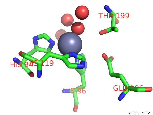
Mono view
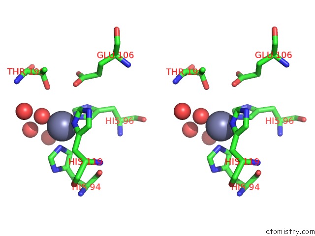
Stereo pair view
Mono view
Stereo pair view
A full contact list of Zinc with other atoms in the Zn binding
site number 1 of X-Ray Crystal Structure of Mutant N62T of Human Carbonic Anhydrase II within 5.0Å range:
|
Reference:
J.Zheng,
B.S.Avvaru,
C.Tu,
R.Mckenna,
D.N.Silverman.
Role of Hydrophilic Residues in Proton Transfer During Catalysis By Human Carbonic Anhydrase II. Biochemistry V. 47 12028 2008.
ISSN: ISSN 0006-2960
PubMed: 18942852
DOI: 10.1021/BI801473W
Page generated: Thu Oct 24 12:20:56 2024
ISSN: ISSN 0006-2960
PubMed: 18942852
DOI: 10.1021/BI801473W
Last articles
Zn in 9MJ5Zn in 9HNW
Zn in 9G0L
Zn in 9FNE
Zn in 9DZN
Zn in 9E0I
Zn in 9D32
Zn in 9DAK
Zn in 8ZXC
Zn in 8ZUF