Zinc »
PDB 2zep-2zxg »
2zog »
Zinc in PDB 2zog: Crystal Structure of Mouse Carnosinase CN2 Complexed with Zn and Bestatin
Enzymatic activity of Crystal Structure of Mouse Carnosinase CN2 Complexed with Zn and Bestatin
All present enzymatic activity of Crystal Structure of Mouse Carnosinase CN2 Complexed with Zn and Bestatin:
3.4.13.18;
3.4.13.18;
Protein crystallography data
The structure of Crystal Structure of Mouse Carnosinase CN2 Complexed with Zn and Bestatin, PDB code: 2zog
was solved by
H.Unno,
T.Yamashita,
N.Okumura,
M.Kusunoki,
with X-Ray Crystallography technique. A brief refinement statistics is given in the table below:
| Resolution Low / High (Å) | 39.34 / 1.70 |
| Space group | P 1 21 1 |
| Cell size a, b, c (Å), α, β, γ (°) | 54.417, 199.779, 55.495, 90.00, 118.52, 90.00 |
| R / Rfree (%) | 19.7 / 24 |
Zinc Binding Sites:
The binding sites of Zinc atom in the Crystal Structure of Mouse Carnosinase CN2 Complexed with Zn and Bestatin
(pdb code 2zog). This binding sites where shown within
5.0 Angstroms radius around Zinc atom.
In total 4 binding sites of Zinc where determined in the Crystal Structure of Mouse Carnosinase CN2 Complexed with Zn and Bestatin, PDB code: 2zog:
Jump to Zinc binding site number: 1; 2; 3; 4;
In total 4 binding sites of Zinc where determined in the Crystal Structure of Mouse Carnosinase CN2 Complexed with Zn and Bestatin, PDB code: 2zog:
Jump to Zinc binding site number: 1; 2; 3; 4;
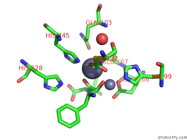
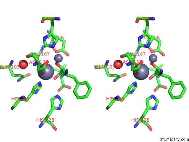
Zinc binding site 1 out of 4 in 2zog
Go back to
Zinc binding site 1 out
of 4 in the Crystal Structure of Mouse Carnosinase CN2 Complexed with Zn and Bestatin
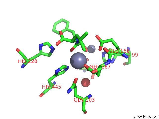
Mono view
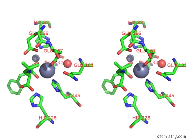
Stereo pair view
Mono view
Stereo pair view
A full contact list of Zinc with other atoms in the Zn binding
site number 1 of Crystal Structure of Mouse Carnosinase CN2 Complexed with Zn and Bestatin within 5.0Å range:
|
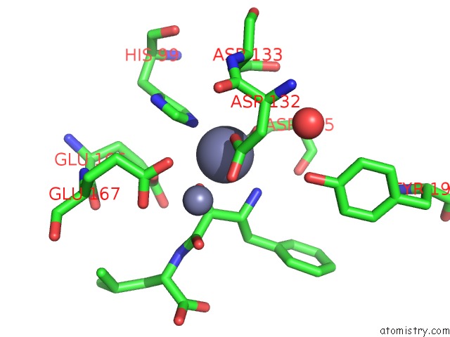
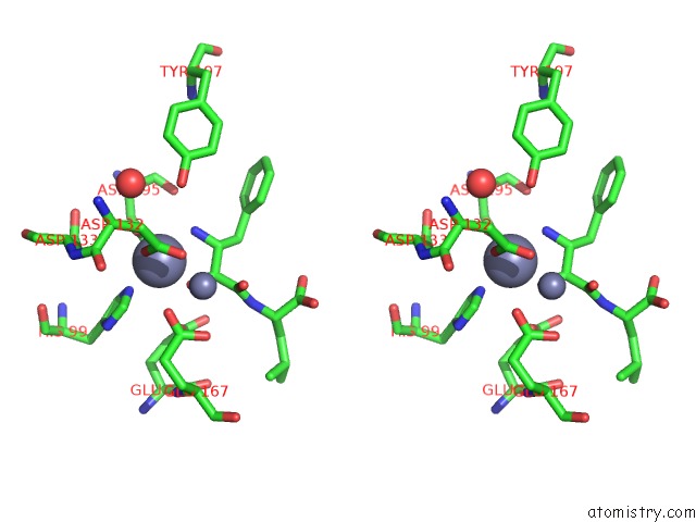
Zinc binding site 2 out of 4 in 2zog
Go back to
Zinc binding site 2 out
of 4 in the Crystal Structure of Mouse Carnosinase CN2 Complexed with Zn and Bestatin
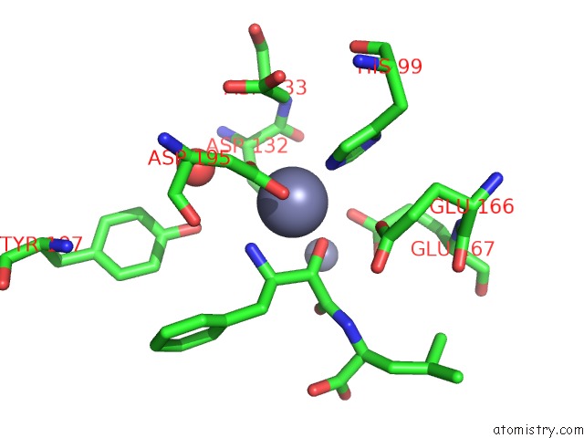
Mono view
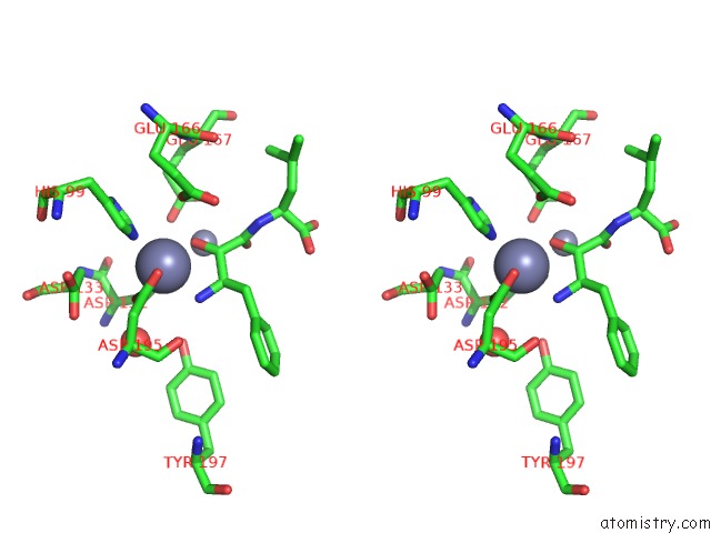
Stereo pair view
Mono view
Stereo pair view
A full contact list of Zinc with other atoms in the Zn binding
site number 2 of Crystal Structure of Mouse Carnosinase CN2 Complexed with Zn and Bestatin within 5.0Å range:
|
Zinc binding site 3 out of 4 in 2zog
Go back to
Zinc binding site 3 out
of 4 in the Crystal Structure of Mouse Carnosinase CN2 Complexed with Zn and Bestatin
Mono view
Stereo pair view
Mono view
Stereo pair view
A full contact list of Zinc with other atoms in the Zn binding
site number 3 of Crystal Structure of Mouse Carnosinase CN2 Complexed with Zn and Bestatin within 5.0Å range:
|
Zinc binding site 4 out of 4 in 2zog
Go back to
Zinc binding site 4 out
of 4 in the Crystal Structure of Mouse Carnosinase CN2 Complexed with Zn and Bestatin
Mono view
Stereo pair view
Mono view
Stereo pair view
A full contact list of Zinc with other atoms in the Zn binding
site number 4 of Crystal Structure of Mouse Carnosinase CN2 Complexed with Zn and Bestatin within 5.0Å range:
|
Reference:
H.Unno,
T.Yamashita,
S.Ujita,
N.Okumura,
H.Otani,
A.Okumura,
K.Nagai,
M.Kusunoki.
Structural Basis For Substrate Recognition and Hydrolysis By Mouse Carnosinase CN2. J.Biol.Chem. V. 283 27289 2008.
ISSN: ISSN 0021-9258
PubMed: 18550540
DOI: 10.1074/JBC.M801657200
Page generated: Thu Oct 24 10:51:28 2024
ISSN: ISSN 0021-9258
PubMed: 18550540
DOI: 10.1074/JBC.M801657200
Last articles
Zn in 9MJ5Zn in 9HNW
Zn in 9G0L
Zn in 9FNE
Zn in 9DZN
Zn in 9E0I
Zn in 9D32
Zn in 9DAK
Zn in 8ZXC
Zn in 8ZUF