Zinc »
PDB 2v20-2vh9 »
2v8d »
Zinc in PDB 2v8d: Crystal Structure of Mutant E159A of Beta-Alanine Synthase From Saccharomyces Kluyveri
Enzymatic activity of Crystal Structure of Mutant E159A of Beta-Alanine Synthase From Saccharomyces Kluyveri
All present enzymatic activity of Crystal Structure of Mutant E159A of Beta-Alanine Synthase From Saccharomyces Kluyveri:
3.5.1.6;
3.5.1.6;
Protein crystallography data
The structure of Crystal Structure of Mutant E159A of Beta-Alanine Synthase From Saccharomyces Kluyveri, PDB code: 2v8d
was solved by
S.Lundgren,
B.Andersen,
J.Piskur,
D.Dobritzsch,
with X-Ray Crystallography technique. A brief refinement statistics is given in the table below:
| Resolution Low / High (Å) | 45.22 / 2.30 |
| Space group | P 1 21 1 |
| Cell size a, b, c (Å), α, β, γ (°) | 61.117, 77.143, 108.228, 90.00, 97.12, 90.00 |
| R / Rfree (%) | 23 / 27.6 |
Zinc Binding Sites:
The binding sites of Zinc atom in the Crystal Structure of Mutant E159A of Beta-Alanine Synthase From Saccharomyces Kluyveri
(pdb code 2v8d). This binding sites where shown within
5.0 Angstroms radius around Zinc atom.
In total 4 binding sites of Zinc where determined in the Crystal Structure of Mutant E159A of Beta-Alanine Synthase From Saccharomyces Kluyveri, PDB code: 2v8d:
Jump to Zinc binding site number: 1; 2; 3; 4;
In total 4 binding sites of Zinc where determined in the Crystal Structure of Mutant E159A of Beta-Alanine Synthase From Saccharomyces Kluyveri, PDB code: 2v8d:
Jump to Zinc binding site number: 1; 2; 3; 4;
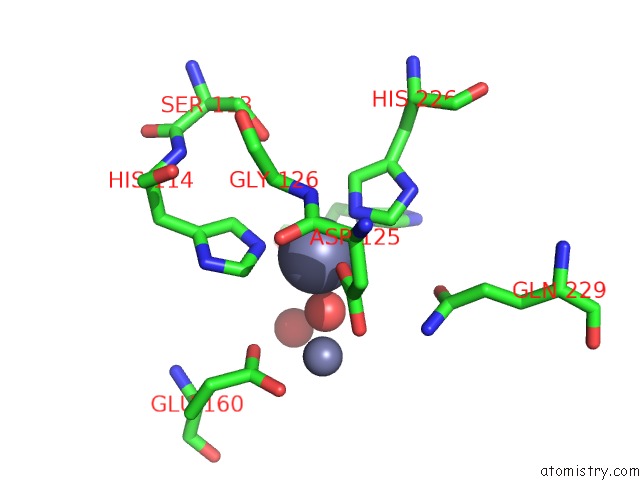
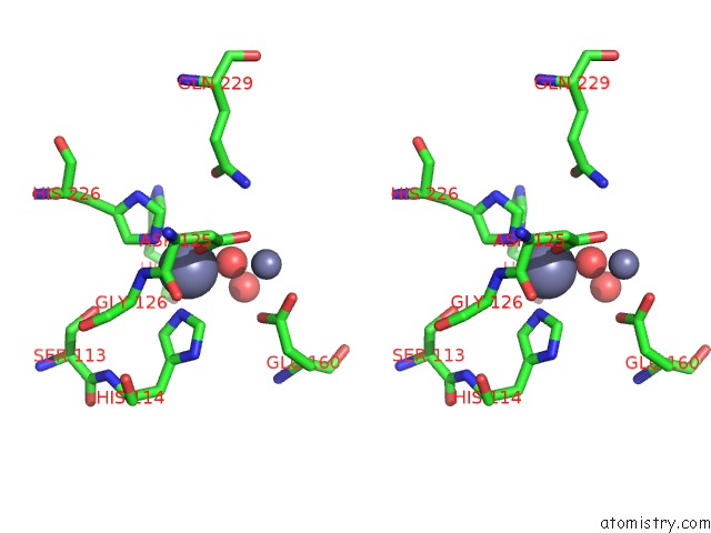
Zinc binding site 1 out of 4 in 2v8d
Go back to
Zinc binding site 1 out
of 4 in the Crystal Structure of Mutant E159A of Beta-Alanine Synthase From Saccharomyces Kluyveri
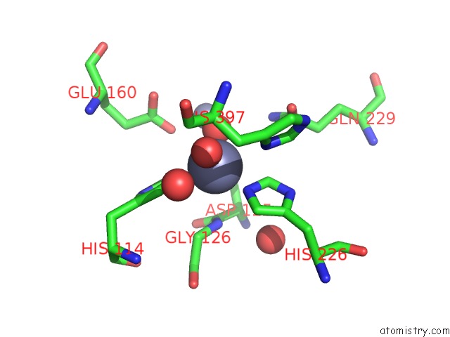
Mono view
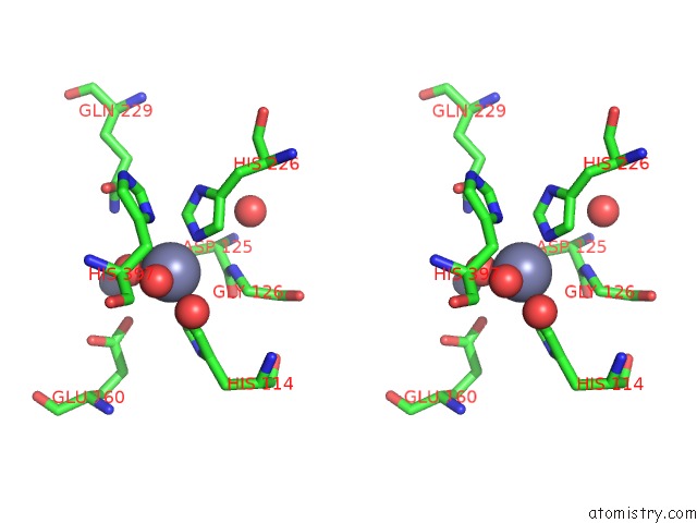
Stereo pair view
Mono view
Stereo pair view
A full contact list of Zinc with other atoms in the Zn binding
site number 1 of Crystal Structure of Mutant E159A of Beta-Alanine Synthase From Saccharomyces Kluyveri within 5.0Å range:
|
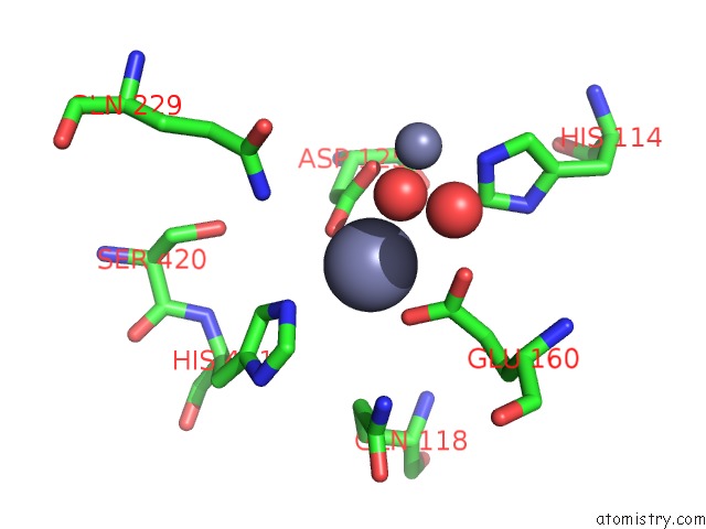
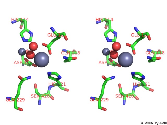
Zinc binding site 2 out of 4 in 2v8d
Go back to
Zinc binding site 2 out
of 4 in the Crystal Structure of Mutant E159A of Beta-Alanine Synthase From Saccharomyces Kluyveri
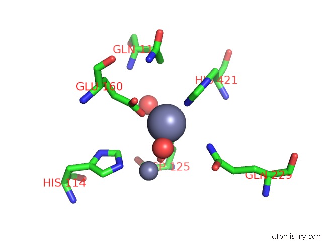
Mono view
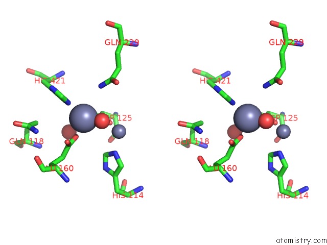
Stereo pair view
Mono view
Stereo pair view
A full contact list of Zinc with other atoms in the Zn binding
site number 2 of Crystal Structure of Mutant E159A of Beta-Alanine Synthase From Saccharomyces Kluyveri within 5.0Å range:
|
Zinc binding site 3 out of 4 in 2v8d
Go back to
Zinc binding site 3 out
of 4 in the Crystal Structure of Mutant E159A of Beta-Alanine Synthase From Saccharomyces Kluyveri
Mono view
Stereo pair view
Mono view
Stereo pair view
A full contact list of Zinc with other atoms in the Zn binding
site number 3 of Crystal Structure of Mutant E159A of Beta-Alanine Synthase From Saccharomyces Kluyveri within 5.0Å range:
|
Zinc binding site 4 out of 4 in 2v8d
Go back to
Zinc binding site 4 out
of 4 in the Crystal Structure of Mutant E159A of Beta-Alanine Synthase From Saccharomyces Kluyveri
Mono view
Stereo pair view
Mono view
Stereo pair view
A full contact list of Zinc with other atoms in the Zn binding
site number 4 of Crystal Structure of Mutant E159A of Beta-Alanine Synthase From Saccharomyces Kluyveri within 5.0Å range:
|
Reference:
S.Lundgren,
B.Andersen,
J.Piskur,
D.Dobritzsch.
Crystal Structures of Yeast -Alanine Synthase Complexes Reveal the Mode of Substrate Binding and Large Scale Domain Closure Movements. J.Biol.Chem. V. 282 36037 2007.
ISSN: ISSN 0021-9258
PubMed: 17916556
DOI: 10.1074/JBC.M705517200
Page generated: Thu Oct 17 04:10:04 2024
ISSN: ISSN 0021-9258
PubMed: 17916556
DOI: 10.1074/JBC.M705517200
Last articles
Zn in 9MJ5Zn in 9HNW
Zn in 9G0L
Zn in 9FNE
Zn in 9DZN
Zn in 9E0I
Zn in 9D32
Zn in 9DAK
Zn in 8ZXC
Zn in 8ZUF