Zinc »
PDB 2q38-2qm1 »
2qiu »
Zinc in PDB 2qiu: Structure of Human Arg-Insulin
Protein crystallography data
The structure of Structure of Human Arg-Insulin, PDB code: 2qiu
was solved by
R.Sreekanth,
V.Pattabhi,
S.S.Rajan,
with X-Ray Crystallography technique. A brief refinement statistics is given in the table below:
| Resolution Low / High (Å) | 40.26 / 2.00 |
| Space group | H 3 |
| Cell size a, b, c (Å), α, β, γ (°) | 80.491, 80.491, 37.640, 90.00, 90.00, 120.00 |
| R / Rfree (%) | 19.7 / 24.9 |
Zinc Binding Sites:
The binding sites of Zinc atom in the Structure of Human Arg-Insulin
(pdb code 2qiu). This binding sites where shown within
5.0 Angstroms radius around Zinc atom.
In total 2 binding sites of Zinc where determined in the Structure of Human Arg-Insulin, PDB code: 2qiu:
Jump to Zinc binding site number: 1; 2;
In total 2 binding sites of Zinc where determined in the Structure of Human Arg-Insulin, PDB code: 2qiu:
Jump to Zinc binding site number: 1; 2;
Zinc binding site 1 out of 2 in 2qiu
Go back to
Zinc binding site 1 out
of 2 in the Structure of Human Arg-Insulin
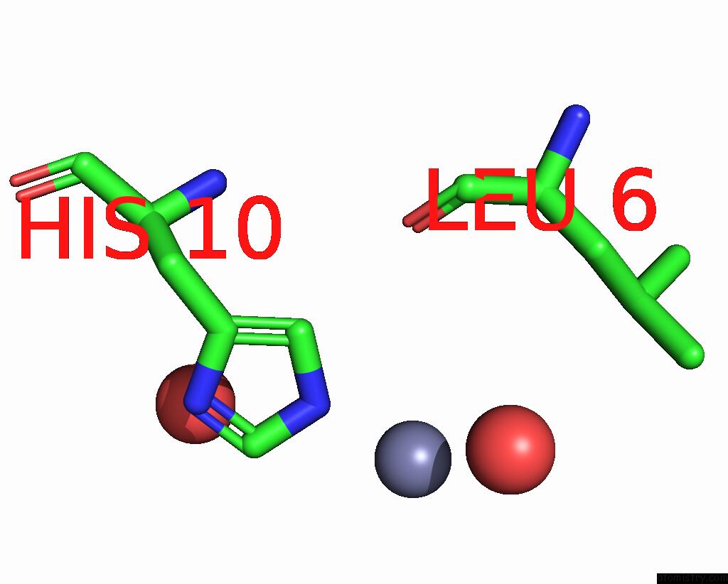
Mono view
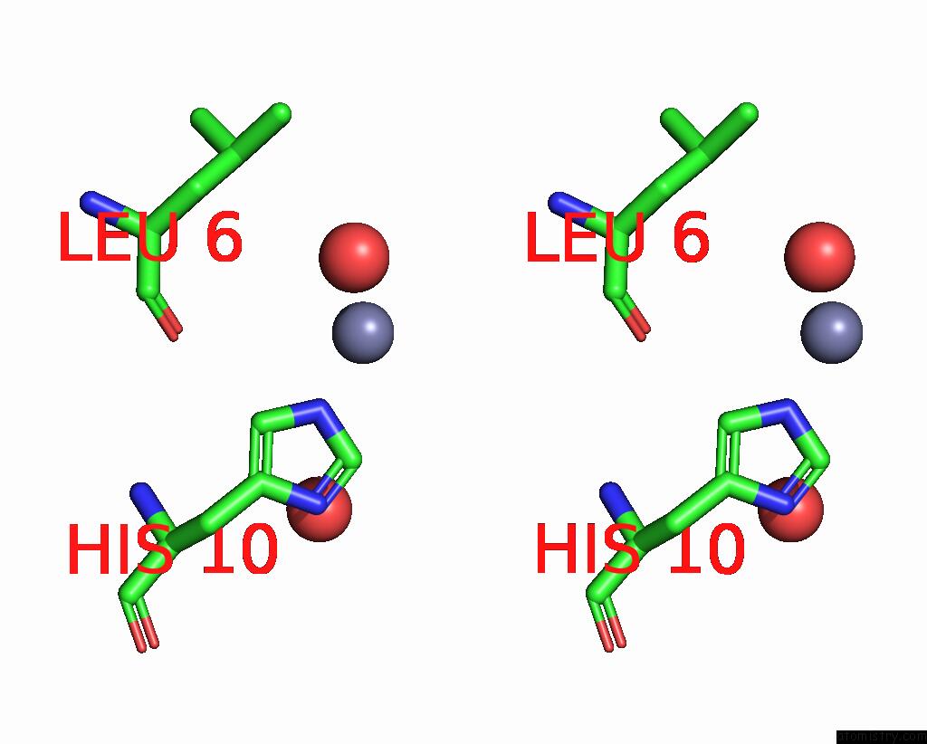
Stereo pair view
Mono view
Stereo pair view
A full contact list of Zinc with other atoms in the Zn binding
site number 1 of Structure of Human Arg-Insulin within 5.0Å range:
|
Zinc binding site 2 out of 2 in 2qiu
Go back to
Zinc binding site 2 out
of 2 in the Structure of Human Arg-Insulin
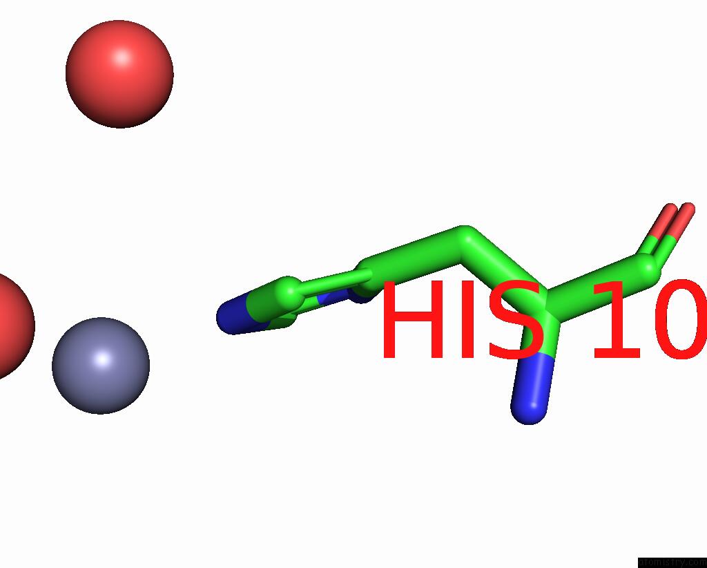
Mono view
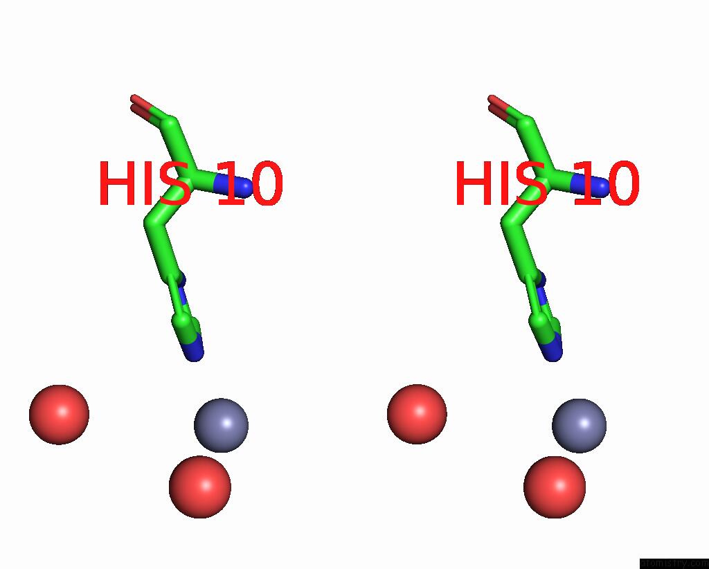
Stereo pair view
Mono view
Stereo pair view
A full contact list of Zinc with other atoms in the Zn binding
site number 2 of Structure of Human Arg-Insulin within 5.0Å range:
|
Reference:
R.Sreekanth,
V.Pattabhi,
S.S.Rajan.
Structural Interpretation of Reduced Insulin Activity As Seen in the Crystal Structure of Human Arg-Insulin Biochimie V. 90 467 2008.
ISSN: ISSN 0300-9084
PubMed: 18029081
DOI: 10.1016/J.BIOCHI.2007.09.012
Page generated: Thu Oct 17 03:28:27 2024
ISSN: ISSN 0300-9084
PubMed: 18029081
DOI: 10.1016/J.BIOCHI.2007.09.012
Last articles
Zn in 9MJ5Zn in 9HNW
Zn in 9G0L
Zn in 9FNE
Zn in 9DZN
Zn in 9E0I
Zn in 9D32
Zn in 9DAK
Zn in 8ZXC
Zn in 8ZUF