Zinc »
PDB 2omi-2ovz »
2osv »
Zinc in PDB 2osv: Crystal Structure of Znua From E. Coli
Protein crystallography data
The structure of Crystal Structure of Znua From E. Coli, PDB code: 2osv
was solved by
H.Li,
G.Jogl,
with X-Ray Crystallography technique. A brief refinement statistics is given in the table below:
| Resolution Low / High (Å) | 30.00 / 1.75 |
| Space group | P 21 21 21 |
| Cell size a, b, c (Å), α, β, γ (°) | 72.980, 87.985, 86.728, 90.00, 90.00, 90.00 |
| R / Rfree (%) | 17.5 / 21.6 |
Zinc Binding Sites:
The binding sites of Zinc atom in the Crystal Structure of Znua From E. Coli
(pdb code 2osv). This binding sites where shown within
5.0 Angstroms radius around Zinc atom.
In total 2 binding sites of Zinc where determined in the Crystal Structure of Znua From E. Coli, PDB code: 2osv:
Jump to Zinc binding site number: 1; 2;
In total 2 binding sites of Zinc where determined in the Crystal Structure of Znua From E. Coli, PDB code: 2osv:
Jump to Zinc binding site number: 1; 2;
Zinc binding site 1 out of 2 in 2osv
Go back to
Zinc binding site 1 out
of 2 in the Crystal Structure of Znua From E. Coli
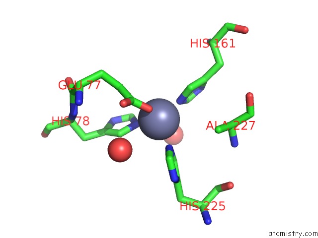
Mono view
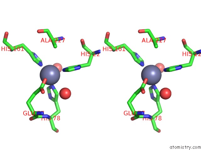
Stereo pair view
Mono view
Stereo pair view
|
|
A full contact list of Zinc with other atoms in the Zn binding
site number 1 of Crystal Structure of Znua From E. Coli within 5.0Å range:
|
Zinc binding site 2 out of 2 in 2osv
Go back to
Zinc binding site 2 out
of 2 in the Crystal Structure of Znua From E. Coli
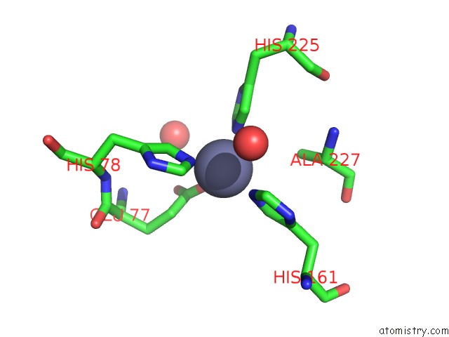
Mono view
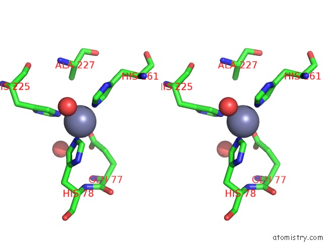
Stereo pair view
Mono view
Stereo pair view
|
|
A full contact list of Zinc with other atoms in the Zn binding
site number 2 of Crystal Structure of Znua From E. Coli within 5.0Å range:
|
Reference:
H.Li,
G.Jogl.
Crystal Structure of the Zinc-Binding Transport Protein Znua From Escherichia Coli Reveals An Unexpected Variation in Metal Coordination. J.Mol.Biol. V. 368 1358 2007.
ISSN: ISSN 0022-2836
PubMed: 17399739
DOI: 10.1016/J.JMB.2007.02.107
Page generated: Thu Oct 17 02:46:04 2024
ISSN: ISSN 0022-2836
PubMed: 17399739
DOI: 10.1016/J.JMB.2007.02.107
Last articles
Zn in 9MJ5Zn in 9HNW
Zn in 9G0L
Zn in 9FNE
Zn in 9DZN
Zn in 9E0I
Zn in 9D32
Zn in 9DAK
Zn in 8ZXC
Zn in 8ZUF