Zinc »
PDB 2nwz-2o6p »
2o4z »
Zinc in PDB 2o4z: Crystal Structure of the Carbonic Anhydrase II Complexed with Hydroxysulfamide Inhibitor
Enzymatic activity of Crystal Structure of the Carbonic Anhydrase II Complexed with Hydroxysulfamide Inhibitor
All present enzymatic activity of Crystal Structure of the Carbonic Anhydrase II Complexed with Hydroxysulfamide Inhibitor:
4.2.1.1;
4.2.1.1;
Protein crystallography data
The structure of Crystal Structure of the Carbonic Anhydrase II Complexed with Hydroxysulfamide Inhibitor, PDB code: 2o4z
was solved by
C.Temperini,
J.Y.Winum,
J.L.Montero,
A.Scozzafava,
C.T.Supuran,
with X-Ray Crystallography technique. A brief refinement statistics is given in the table below:
| Resolution Low / High (Å) | 20.00 / 2.10 |
| Space group | P 1 21 1 |
| Cell size a, b, c (Å), α, β, γ (°) | 42.100, 41.390, 72.300, 90.00, 104.38, 90.00 |
| R / Rfree (%) | 20.8 / 26.6 |
Other elements in 2o4z:
The structure of Crystal Structure of the Carbonic Anhydrase II Complexed with Hydroxysulfamide Inhibitor also contains other interesting chemical elements:
| Mercury | (Hg) | 1 atom |
Zinc Binding Sites:
The binding sites of Zinc atom in the Crystal Structure of the Carbonic Anhydrase II Complexed with Hydroxysulfamide Inhibitor
(pdb code 2o4z). This binding sites where shown within
5.0 Angstroms radius around Zinc atom.
In total only one binding site of Zinc was determined in the Crystal Structure of the Carbonic Anhydrase II Complexed with Hydroxysulfamide Inhibitor, PDB code: 2o4z:
In total only one binding site of Zinc was determined in the Crystal Structure of the Carbonic Anhydrase II Complexed with Hydroxysulfamide Inhibitor, PDB code: 2o4z:
Zinc binding site 1 out of 1 in 2o4z
Go back to
Zinc binding site 1 out
of 1 in the Crystal Structure of the Carbonic Anhydrase II Complexed with Hydroxysulfamide Inhibitor
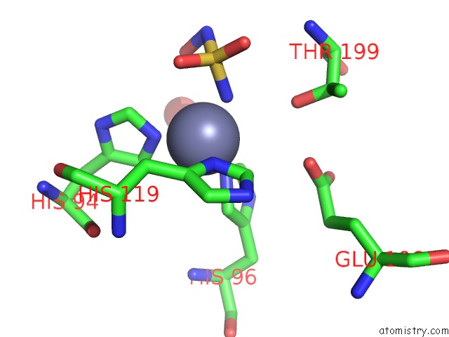
Mono view
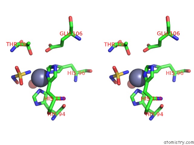
Stereo pair view
Mono view
Stereo pair view
A full contact list of Zinc with other atoms in the Zn binding
site number 1 of Crystal Structure of the Carbonic Anhydrase II Complexed with Hydroxysulfamide Inhibitor within 5.0Å range:
|
Reference:
C.Temperini,
J.Y.Winum,
J.L.Montero,
A.Scozzafava,
C.T.Supuran.
Carbonic Anhydrase Inhibitors: the X-Ray Crystal Structure of the Adduct of N-Hydroxysulfamide with Isozyme II Explains Why This New Zinc Binding Function Is Effective in the Design of Potent Inhibitors. Bioorg.Med.Chem.Lett. V. 17 2795 2007.
ISSN: ISSN 0960-894X
PubMed: 17346964
DOI: 10.1016/J.BMCL.2007.02.068
Page generated: Thu Oct 17 02:27:06 2024
ISSN: ISSN 0960-894X
PubMed: 17346964
DOI: 10.1016/J.BMCL.2007.02.068
Last articles
Zn in 9MJ5Zn in 9HNW
Zn in 9G0L
Zn in 9FNE
Zn in 9DZN
Zn in 9E0I
Zn in 9D32
Zn in 9DAK
Zn in 8ZXC
Zn in 8ZUF