Zinc »
PDB 2naa-2nwy »
2nso »
Zinc in PDB 2nso: Trna-Gunanine-Transglycosylase (Tgt) Mutant Y106F, C158V, A232S, V233G- Apo-Structure
Enzymatic activity of Trna-Gunanine-Transglycosylase (Tgt) Mutant Y106F, C158V, A232S, V233G- Apo-Structure
All present enzymatic activity of Trna-Gunanine-Transglycosylase (Tgt) Mutant Y106F, C158V, A232S, V233G- Apo-Structure:
2.4.2.29;
2.4.2.29;
Protein crystallography data
The structure of Trna-Gunanine-Transglycosylase (Tgt) Mutant Y106F, C158V, A232S, V233G- Apo-Structure, PDB code: 2nso
was solved by
N.Tidten,
A.Heine,
K.Reuter,
G.Klebe,
with X-Ray Crystallography technique. A brief refinement statistics is given in the table below:
| Resolution Low / High (Å) | 28.42 / 1.60 |
| Space group | C 1 2 1 |
| Cell size a, b, c (Å), α, β, γ (°) | 90.730, 64.830, 70.810, 90.00, 96.31, 90.00 |
| R / Rfree (%) | 16.2 / 22.1 |
Zinc Binding Sites:
The binding sites of Zinc atom in the Trna-Gunanine-Transglycosylase (Tgt) Mutant Y106F, C158V, A232S, V233G- Apo-Structure
(pdb code 2nso). This binding sites where shown within
5.0 Angstroms radius around Zinc atom.
In total only one binding site of Zinc was determined in the Trna-Gunanine-Transglycosylase (Tgt) Mutant Y106F, C158V, A232S, V233G- Apo-Structure, PDB code: 2nso:
In total only one binding site of Zinc was determined in the Trna-Gunanine-Transglycosylase (Tgt) Mutant Y106F, C158V, A232S, V233G- Apo-Structure, PDB code: 2nso:
Zinc binding site 1 out of 1 in 2nso
Go back to
Zinc binding site 1 out
of 1 in the Trna-Gunanine-Transglycosylase (Tgt) Mutant Y106F, C158V, A232S, V233G- Apo-Structure
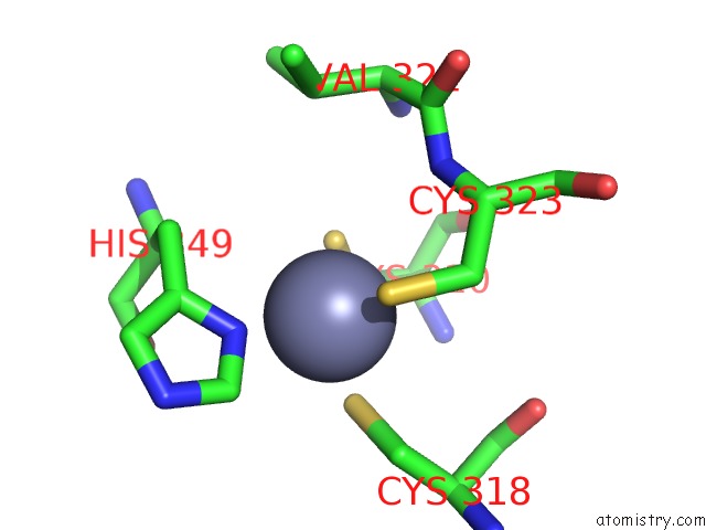
Mono view
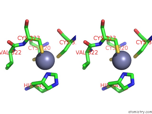
Stereo pair view
Mono view
Stereo pair view
A full contact list of Zinc with other atoms in the Zn binding
site number 1 of Trna-Gunanine-Transglycosylase (Tgt) Mutant Y106F, C158V, A232S, V233G- Apo-Structure within 5.0Å range:
|
Reference:
I.Biela,
N.Tidten-Luksch,
F.Immekus,
S.Glinca,
T.X.Nguyen,
H.D.Gerber,
A.Heine,
G.Klebe,
K.Reuter.
Investigation of Specificity Determinants in Bacterial Trna-Guanine Transglycosylase Reveals Queuine, the Substrate of Its Eucaryotic Counterpart, As Inhibitor. Plos One V. 8 64240 2013.
ISSN: ESSN 1932-6203
PubMed: 23704982
DOI: 10.1371/JOURNAL.PONE.0064240
Page generated: Thu Oct 17 02:17:18 2024
ISSN: ESSN 1932-6203
PubMed: 23704982
DOI: 10.1371/JOURNAL.PONE.0064240
Last articles
Zn in 9MJ5Zn in 9HNW
Zn in 9G0L
Zn in 9FNE
Zn in 9DZN
Zn in 9E0I
Zn in 9D32
Zn in 9DAK
Zn in 8ZXC
Zn in 8ZUF