Zinc »
PDB 2naa-2nwy »
2nqj »
Zinc in PDB 2nqj: Crystal Structure of Escherichia Coli Endonuclease IV (Endo IV) E261Q Mutant Bound to Damaged Dna
Enzymatic activity of Crystal Structure of Escherichia Coli Endonuclease IV (Endo IV) E261Q Mutant Bound to Damaged Dna
All present enzymatic activity of Crystal Structure of Escherichia Coli Endonuclease IV (Endo IV) E261Q Mutant Bound to Damaged Dna:
3.1.21.2;
3.1.21.2;
Protein crystallography data
The structure of Crystal Structure of Escherichia Coli Endonuclease IV (Endo IV) E261Q Mutant Bound to Damaged Dna, PDB code: 2nqj
was solved by
E.D.Garcin-Hosfield,
D.J.Hosfield,
J.A.Tainer,
with X-Ray Crystallography technique. A brief refinement statistics is given in the table below:
| Resolution Low / High (Å) | 20.00 / 2.45 |
| Space group | P 21 21 21 |
| Cell size a, b, c (Å), α, β, γ (°) | 55.600, 86.200, 148.300, 90.00, 90.00, 90.00 |
| R / Rfree (%) | 19.8 / 25.4 |
Zinc Binding Sites:
The binding sites of Zinc atom in the Crystal Structure of Escherichia Coli Endonuclease IV (Endo IV) E261Q Mutant Bound to Damaged Dna
(pdb code 2nqj). This binding sites where shown within
5.0 Angstroms radius around Zinc atom.
In total 4 binding sites of Zinc where determined in the Crystal Structure of Escherichia Coli Endonuclease IV (Endo IV) E261Q Mutant Bound to Damaged Dna, PDB code: 2nqj:
Jump to Zinc binding site number: 1; 2; 3; 4;
In total 4 binding sites of Zinc where determined in the Crystal Structure of Escherichia Coli Endonuclease IV (Endo IV) E261Q Mutant Bound to Damaged Dna, PDB code: 2nqj:
Jump to Zinc binding site number: 1; 2; 3; 4;
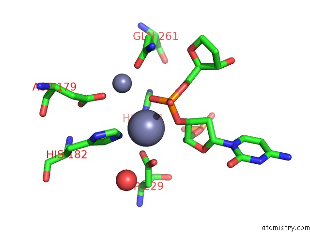
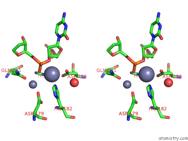
Zinc binding site 1 out of 4 in 2nqj
Go back to
Zinc binding site 1 out
of 4 in the Crystal Structure of Escherichia Coli Endonuclease IV (Endo IV) E261Q Mutant Bound to Damaged Dna
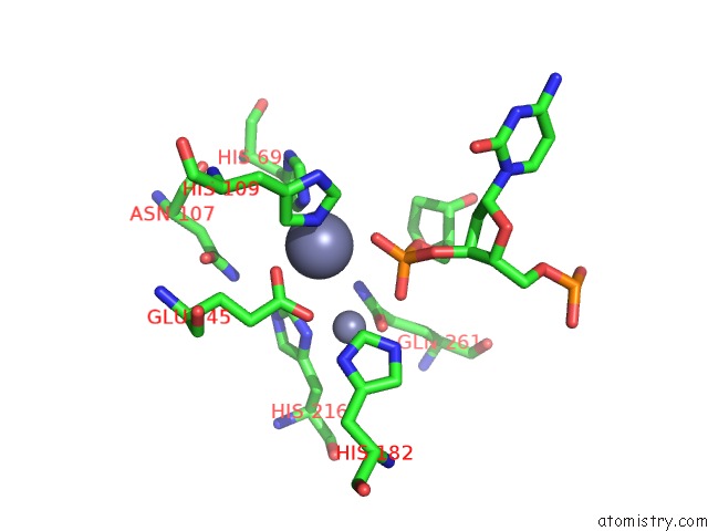
Mono view
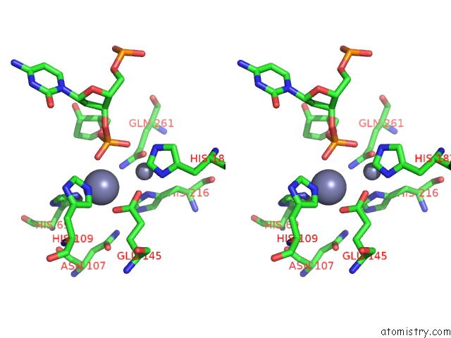
Stereo pair view
Mono view
Stereo pair view
A full contact list of Zinc with other atoms in the Zn binding
site number 1 of Crystal Structure of Escherichia Coli Endonuclease IV (Endo IV) E261Q Mutant Bound to Damaged Dna within 5.0Å range:
|
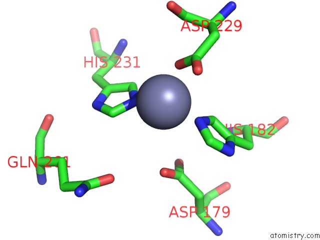
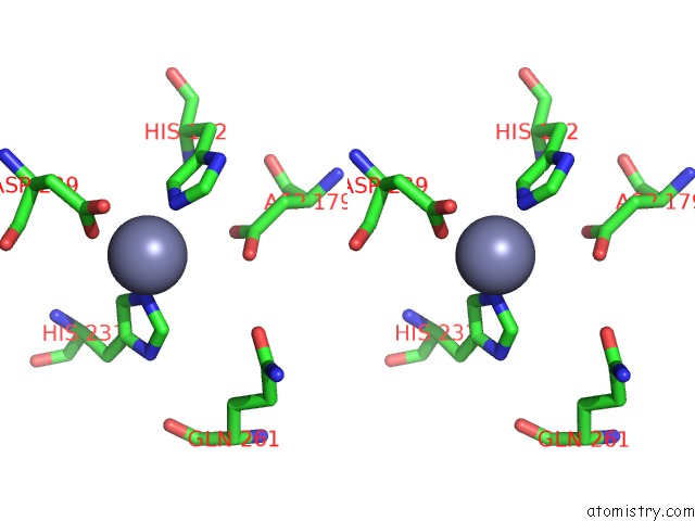
Zinc binding site 2 out of 4 in 2nqj
Go back to
Zinc binding site 2 out
of 4 in the Crystal Structure of Escherichia Coli Endonuclease IV (Endo IV) E261Q Mutant Bound to Damaged Dna
Mono view
Stereo pair view
Mono view
Stereo pair view
A full contact list of Zinc with other atoms in the Zn binding
site number 2 of Crystal Structure of Escherichia Coli Endonuclease IV (Endo IV) E261Q Mutant Bound to Damaged Dna within 5.0Å range:
|
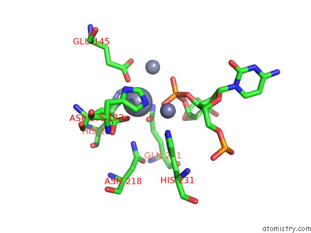
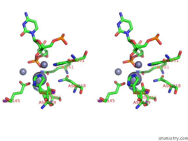
Zinc binding site 3 out of 4 in 2nqj
Go back to
Zinc binding site 3 out
of 4 in the Crystal Structure of Escherichia Coli Endonuclease IV (Endo IV) E261Q Mutant Bound to Damaged Dna
Mono view
Stereo pair view
Mono view
Stereo pair view
A full contact list of Zinc with other atoms in the Zn binding
site number 3 of Crystal Structure of Escherichia Coli Endonuclease IV (Endo IV) E261Q Mutant Bound to Damaged Dna within 5.0Å range:
|
Zinc binding site 4 out of 4 in 2nqj
Go back to
Zinc binding site 4 out
of 4 in the Crystal Structure of Escherichia Coli Endonuclease IV (Endo IV) E261Q Mutant Bound to Damaged Dna
Mono view
Stereo pair view
Mono view
Stereo pair view
A full contact list of Zinc with other atoms in the Zn binding
site number 4 of Crystal Structure of Escherichia Coli Endonuclease IV (Endo IV) E261Q Mutant Bound to Damaged Dna within 5.0Å range:
|
Reference:
E.D.Garcin,
D.J.Hosfield,
S.A.Desai,
B.J.Haas,
M.Bjoras,
R.P.Cunningham,
J.A.Tainer.
Dna Apurinic-Apyrimidinic Site Binding and Excision By Endonuclease IV. Nat.Struct.Mol.Biol. V. 15 515 2008.
ISSN: ISSN 1545-9993
PubMed: 18408731
DOI: 10.1038/NSMB.1414
Page generated: Thu Oct 17 02:16:03 2024
ISSN: ISSN 1545-9993
PubMed: 18408731
DOI: 10.1038/NSMB.1414
Last articles
Zn in 9MJ5Zn in 9HNW
Zn in 9G0L
Zn in 9FNE
Zn in 9DZN
Zn in 9E0I
Zn in 9D32
Zn in 9DAK
Zn in 8ZXC
Zn in 8ZUF