Zinc »
PDB 2naa-2nwy »
2nnv »
Zinc in PDB 2nnv: Structure of Inhibitor Binding to Carbonic Anhydrase II
Enzymatic activity of Structure of Inhibitor Binding to Carbonic Anhydrase II
All present enzymatic activity of Structure of Inhibitor Binding to Carbonic Anhydrase II:
4.2.1.1;
4.2.1.1;
Protein crystallography data
The structure of Structure of Inhibitor Binding to Carbonic Anhydrase II, PDB code: 2nnv
was solved by
D.W.Christianson,
K.M.Jude,
with X-Ray Crystallography technique. A brief refinement statistics is given in the table below:
| Resolution Low / High (Å) | 80.00 / 1.10 |
| Space group | P 1 21 1 |
| Cell size a, b, c (Å), α, β, γ (°) | 42.365, 41.213, 72.145, 90.00, 104.83, 90.00 |
| R / Rfree (%) | 13.2 / 16.4 |
Other elements in 2nnv:
The structure of Structure of Inhibitor Binding to Carbonic Anhydrase II also contains other interesting chemical elements:
| Mercury | (Hg) | 1 atom |
Zinc Binding Sites:
The binding sites of Zinc atom in the Structure of Inhibitor Binding to Carbonic Anhydrase II
(pdb code 2nnv). This binding sites where shown within
5.0 Angstroms radius around Zinc atom.
In total only one binding site of Zinc was determined in the Structure of Inhibitor Binding to Carbonic Anhydrase II, PDB code: 2nnv:
In total only one binding site of Zinc was determined in the Structure of Inhibitor Binding to Carbonic Anhydrase II, PDB code: 2nnv:
Zinc binding site 1 out of 1 in 2nnv
Go back to
Zinc binding site 1 out
of 1 in the Structure of Inhibitor Binding to Carbonic Anhydrase II
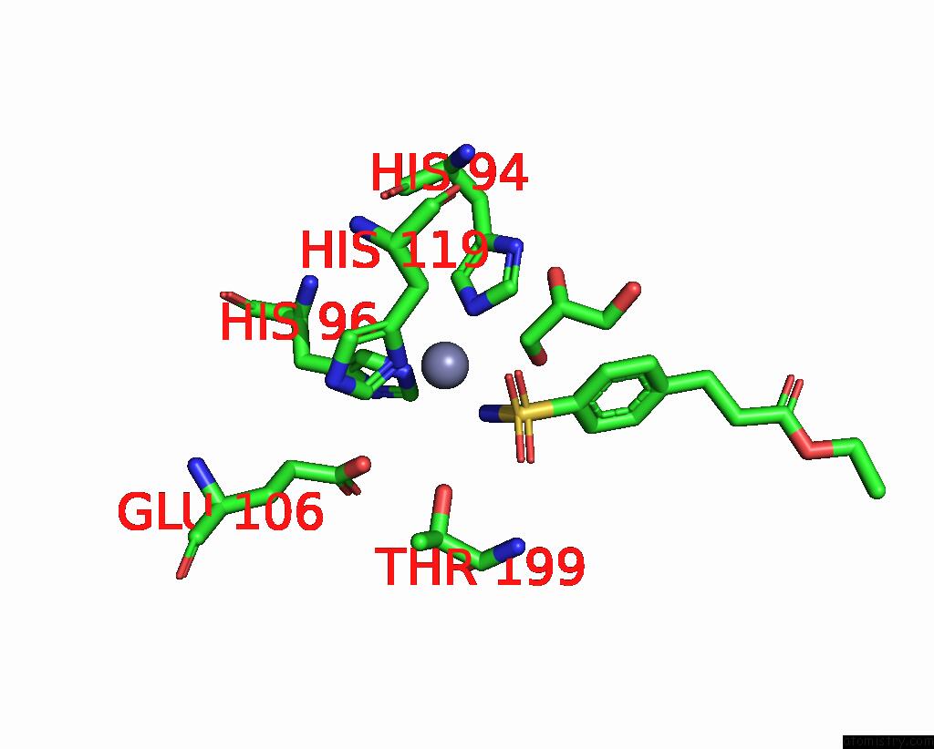
Mono view
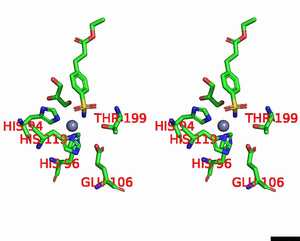
Stereo pair view
Mono view
Stereo pair view
A full contact list of Zinc with other atoms in the Zn binding
site number 1 of Structure of Inhibitor Binding to Carbonic Anhydrase II within 5.0Å range:
|
Reference:
D.K.Srivastava,
K.M.Jude,
A.L.Banerjee,
M.Haldar,
S.Manokaran,
J.Kooren,
S.Mallik,
D.W.Christianson.
Structural Analysis of Charge Discrimination in the Binding of Inhibitors to Human Carbonic Anhydrases I and II. J.Am.Chem.Soc. V. 129 5528 2007.
ISSN: ISSN 0002-7863
PubMed: 17407288
DOI: 10.1021/JA068359W
Page generated: Thu Oct 17 02:15:28 2024
ISSN: ISSN 0002-7863
PubMed: 17407288
DOI: 10.1021/JA068359W
Last articles
Zn in 9MJ5Zn in 9HNW
Zn in 9G0L
Zn in 9FNE
Zn in 9DZN
Zn in 9E0I
Zn in 9D32
Zn in 9DAK
Zn in 8ZXC
Zn in 8ZUF