Zinc »
PDB 2gzi-2han »
2h6t »
Zinc in PDB 2h6t: Secreted Aspartic Proteinase (Sap) 3 From Candida Albicans Complexed with Pepstatin A
Enzymatic activity of Secreted Aspartic Proteinase (Sap) 3 From Candida Albicans Complexed with Pepstatin A
All present enzymatic activity of Secreted Aspartic Proteinase (Sap) 3 From Candida Albicans Complexed with Pepstatin A:
3.4.23.24;
3.4.23.24;
Protein crystallography data
The structure of Secreted Aspartic Proteinase (Sap) 3 From Candida Albicans Complexed with Pepstatin A, PDB code: 2h6t
was solved by
E.Ruge,
C.Borelli,
K.Maskos,
R.Huber,
with X-Ray Crystallography technique. A brief refinement statistics is given in the table below:
| Resolution Low / High (Å) | 19.86 / 1.90 |
| Space group | P 32 2 1 |
| Cell size a, b, c (Å), α, β, γ (°) | 61.100, 61.100, 170.740, 90.00, 90.00, 120.00 |
| R / Rfree (%) | 23.9 / 25.1 |
Zinc Binding Sites:
The binding sites of Zinc atom in the Secreted Aspartic Proteinase (Sap) 3 From Candida Albicans Complexed with Pepstatin A
(pdb code 2h6t). This binding sites where shown within
5.0 Angstroms radius around Zinc atom.
In total only one binding site of Zinc was determined in the Secreted Aspartic Proteinase (Sap) 3 From Candida Albicans Complexed with Pepstatin A, PDB code: 2h6t:
In total only one binding site of Zinc was determined in the Secreted Aspartic Proteinase (Sap) 3 From Candida Albicans Complexed with Pepstatin A, PDB code: 2h6t:
Zinc binding site 1 out of 1 in 2h6t
Go back to
Zinc binding site 1 out
of 1 in the Secreted Aspartic Proteinase (Sap) 3 From Candida Albicans Complexed with Pepstatin A
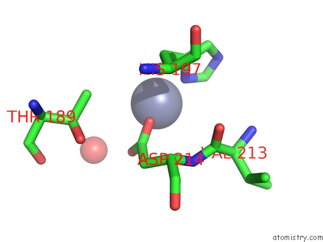
Mono view
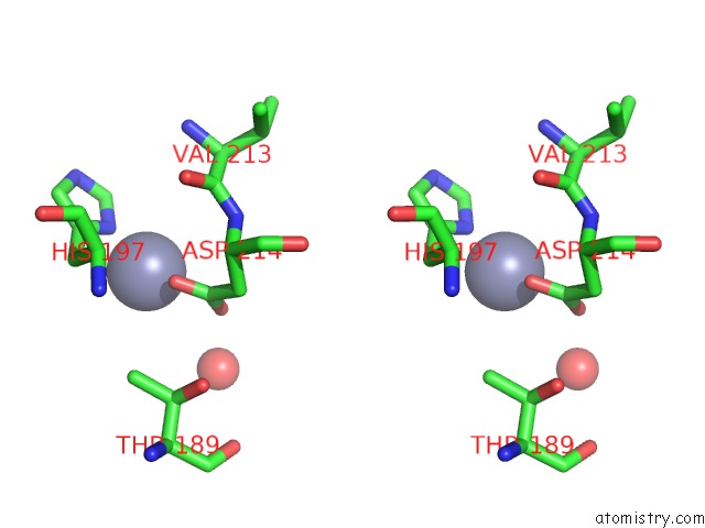
Stereo pair view
Mono view
Stereo pair view
A full contact list of Zinc with other atoms in the Zn binding
site number 1 of Secreted Aspartic Proteinase (Sap) 3 From Candida Albicans Complexed with Pepstatin A within 5.0Å range:
|
Reference:
C.Borelli,
E.Ruge,
M.Schaller,
M.Monod,
H.C.Korting,
R.Huber,
K.Maskos.
The Crystal Structure of the Secreted Aspartic Proteinase 3 From Candida Albicans and Its Complex with Pepstatin A. Proteins V. 68 738 2007.
ISSN: ISSN 0887-3585
PubMed: 17510964
DOI: 10.1002/PROT.21425
Page generated: Thu Oct 17 00:33:20 2024
ISSN: ISSN 0887-3585
PubMed: 17510964
DOI: 10.1002/PROT.21425
Last articles
Zn in 9MJ5Zn in 9HNW
Zn in 9G0L
Zn in 9FNE
Zn in 9DZN
Zn in 9E0I
Zn in 9D32
Zn in 9DAK
Zn in 8ZXC
Zn in 8ZUF