Zinc »
PDB 2eex-2elu »
2eg7 »
Zinc in PDB 2eg7: The Crystal Structure of E. Coli Dihydroorotase Complexed with Hddp
Enzymatic activity of The Crystal Structure of E. Coli Dihydroorotase Complexed with Hddp
All present enzymatic activity of The Crystal Structure of E. Coli Dihydroorotase Complexed with Hddp:
3.5.2.3;
3.5.2.3;
Protein crystallography data
The structure of The Crystal Structure of E. Coli Dihydroorotase Complexed with Hddp, PDB code: 2eg7
was solved by
M.Lee,
M.J.Maher,
J.M.Guss,
with X-Ray Crystallography technique. A brief refinement statistics is given in the table below:
| Resolution Low / High (Å) | 30.00 / 2.00 |
| Space group | P 21 21 21 |
| Cell size a, b, c (Å), α, β, γ (°) | 51.585, 79.628, 180.652, 90.00, 90.00, 90.00 |
| R / Rfree (%) | 18 / 23.5 |
Zinc Binding Sites:
The binding sites of Zinc atom in the The Crystal Structure of E. Coli Dihydroorotase Complexed with Hddp
(pdb code 2eg7). This binding sites where shown within
5.0 Angstroms radius around Zinc atom.
In total 4 binding sites of Zinc where determined in the The Crystal Structure of E. Coli Dihydroorotase Complexed with Hddp, PDB code: 2eg7:
Jump to Zinc binding site number: 1; 2; 3; 4;
In total 4 binding sites of Zinc where determined in the The Crystal Structure of E. Coli Dihydroorotase Complexed with Hddp, PDB code: 2eg7:
Jump to Zinc binding site number: 1; 2; 3; 4;
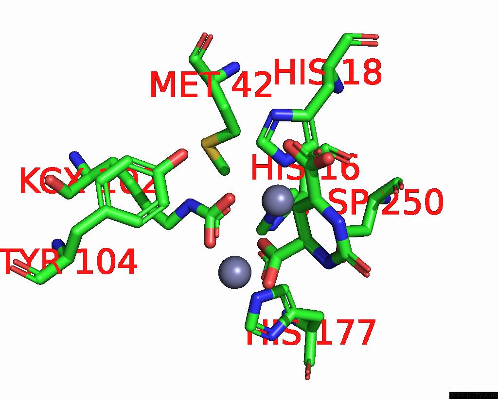
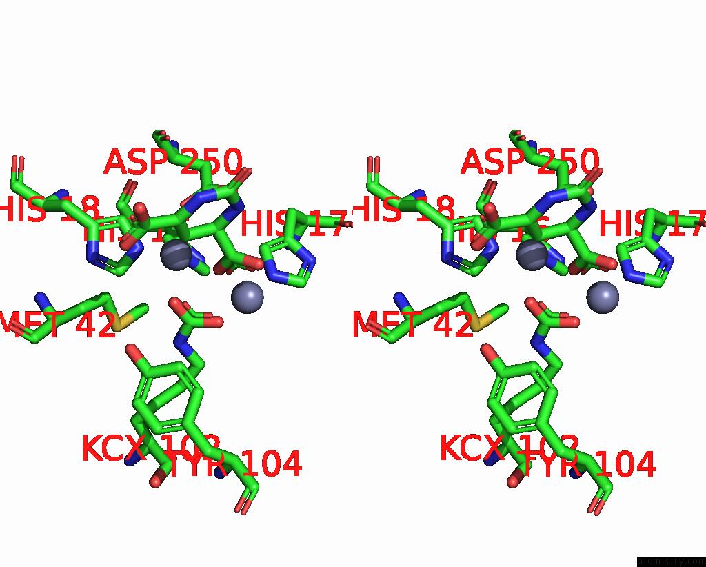
Zinc binding site 1 out of 4 in 2eg7
Go back to
Zinc binding site 1 out
of 4 in the The Crystal Structure of E. Coli Dihydroorotase Complexed with Hddp
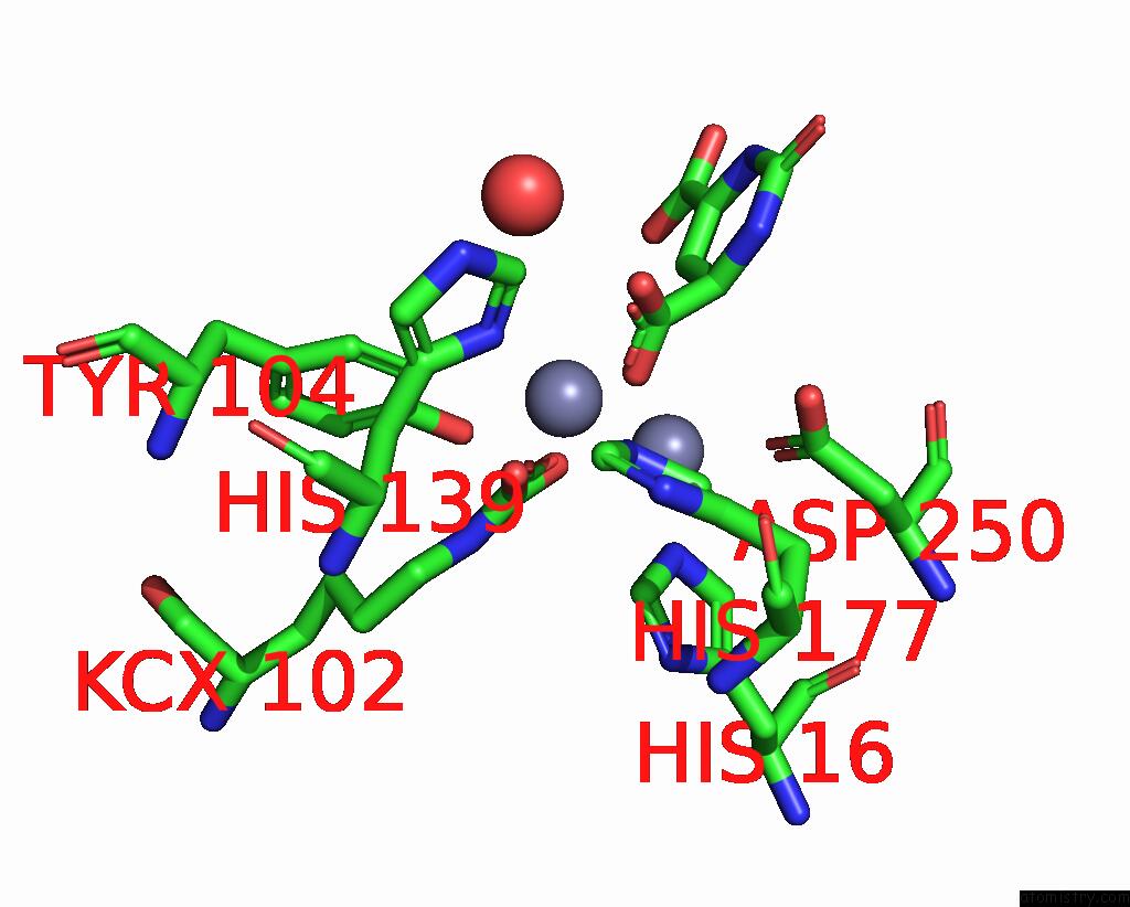
Mono view
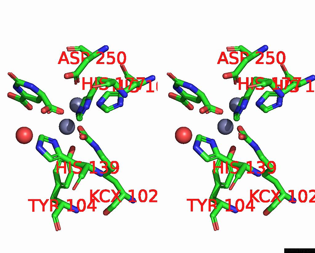
Stereo pair view
Mono view
Stereo pair view
A full contact list of Zinc with other atoms in the Zn binding
site number 1 of The Crystal Structure of E. Coli Dihydroorotase Complexed with Hddp within 5.0Å range:
|
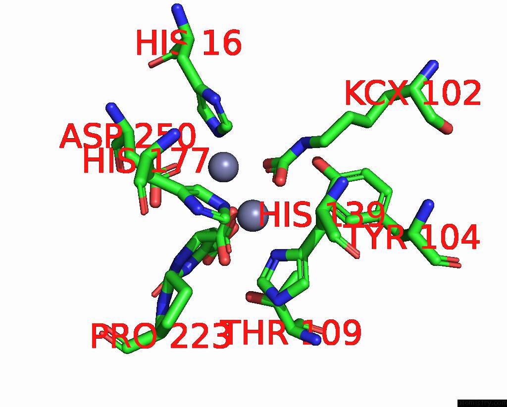
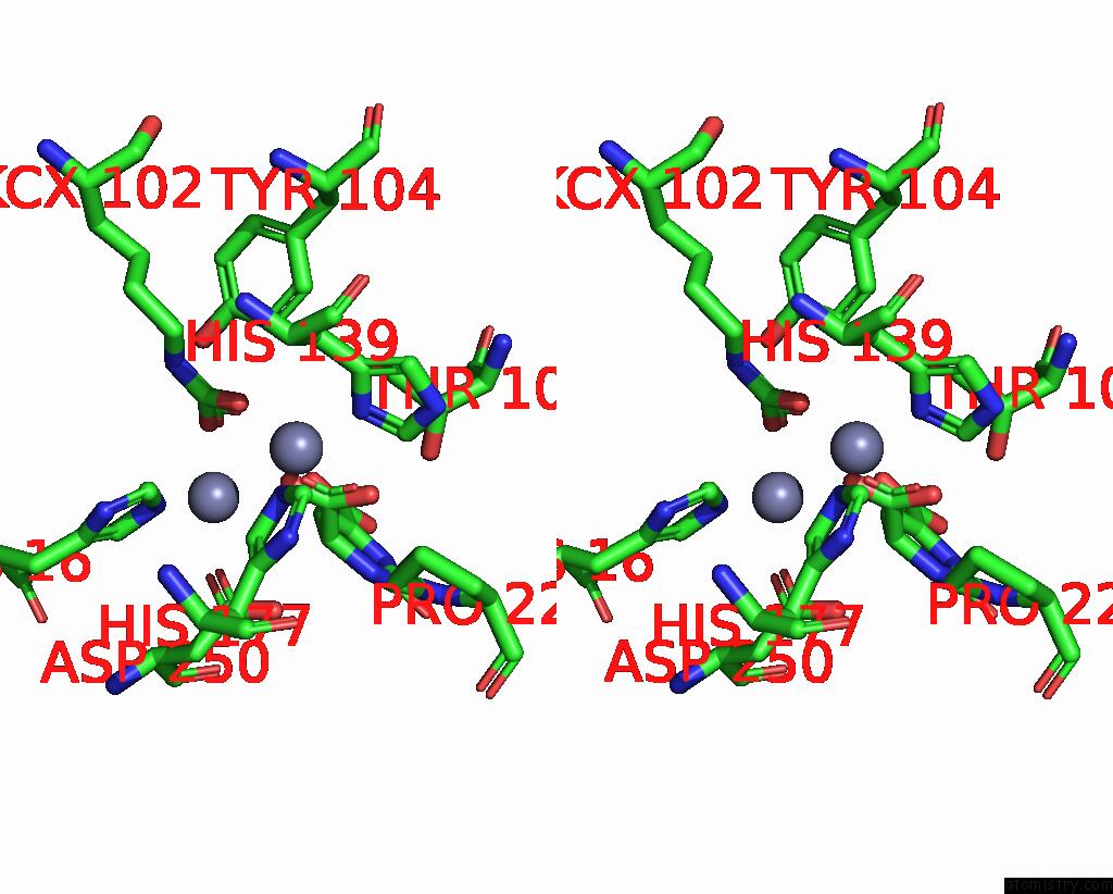
Zinc binding site 2 out of 4 in 2eg7
Go back to
Zinc binding site 2 out
of 4 in the The Crystal Structure of E. Coli Dihydroorotase Complexed with Hddp
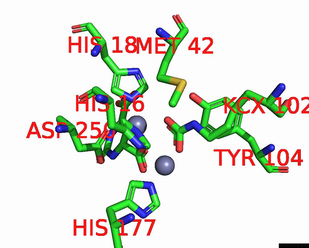
Mono view
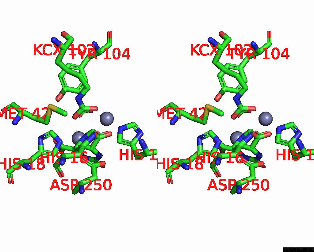
Stereo pair view
Mono view
Stereo pair view
A full contact list of Zinc with other atoms in the Zn binding
site number 2 of The Crystal Structure of E. Coli Dihydroorotase Complexed with Hddp within 5.0Å range:
|
Zinc binding site 3 out of 4 in 2eg7
Go back to
Zinc binding site 3 out
of 4 in the The Crystal Structure of E. Coli Dihydroorotase Complexed with Hddp
Mono view
Stereo pair view
Mono view
Stereo pair view
A full contact list of Zinc with other atoms in the Zn binding
site number 3 of The Crystal Structure of E. Coli Dihydroorotase Complexed with Hddp within 5.0Å range:
|
Zinc binding site 4 out of 4 in 2eg7
Go back to
Zinc binding site 4 out
of 4 in the The Crystal Structure of E. Coli Dihydroorotase Complexed with Hddp
Mono view
Stereo pair view
Mono view
Stereo pair view
A full contact list of Zinc with other atoms in the Zn binding
site number 4 of The Crystal Structure of E. Coli Dihydroorotase Complexed with Hddp within 5.0Å range:
|
Reference:
M.Lee,
C.W.Chan,
S.C.Graham,
R.I.Christopherson,
J.M.Guss,
M.J.Maher.
Structures of Ligand-Free and Inhibitor Complexes of Dihydroorotase From Escherichia Coli: Implications For Loop Movement in Inhibitor Design J.Mol.Biol. V. 370 812 2007.
ISSN: ISSN 0022-2836
PubMed: 17550785
DOI: 10.1016/J.JMB.2007.05.019
Page generated: Wed Oct 16 23:07:13 2024
ISSN: ISSN 0022-2836
PubMed: 17550785
DOI: 10.1016/J.JMB.2007.05.019
Last articles
Zn in 9MJ5Zn in 9HNW
Zn in 9G0L
Zn in 9FNE
Zn in 9DZN
Zn in 9E0I
Zn in 9D32
Zn in 9DAK
Zn in 8ZXC
Zn in 8ZUF