Zinc »
PDB 1zfp-1zsb »
1zli »
Zinc in PDB 1zli: Crystal Structure of the Tick Carboxypeptidase Inhibitor in Complex with Human Carboxypeptidase B
Enzymatic activity of Crystal Structure of the Tick Carboxypeptidase Inhibitor in Complex with Human Carboxypeptidase B
All present enzymatic activity of Crystal Structure of the Tick Carboxypeptidase Inhibitor in Complex with Human Carboxypeptidase B:
3.4.17.2;
3.4.17.2;
Protein crystallography data
The structure of Crystal Structure of the Tick Carboxypeptidase Inhibitor in Complex with Human Carboxypeptidase B, PDB code: 1zli
was solved by
J.L.Arolas,
G.M.Popowicz,
J.Lorenzo,
C.P.Sommerhoff,
R.Huber,
F.X.Aviles,
T.A.Holak,
with X-Ray Crystallography technique. A brief refinement statistics is given in the table below:
| Resolution Low / High (Å) | 19.96 / 2.09 |
| Space group | P 43 21 2 |
| Cell size a, b, c (Å), α, β, γ (°) | 74.200, 74.200, 163.550, 90.00, 90.00, 90.00 |
| R / Rfree (%) | 15.7 / 21.4 |
Zinc Binding Sites:
The binding sites of Zinc atom in the Crystal Structure of the Tick Carboxypeptidase Inhibitor in Complex with Human Carboxypeptidase B
(pdb code 1zli). This binding sites where shown within
5.0 Angstroms radius around Zinc atom.
In total only one binding site of Zinc was determined in the Crystal Structure of the Tick Carboxypeptidase Inhibitor in Complex with Human Carboxypeptidase B, PDB code: 1zli:
In total only one binding site of Zinc was determined in the Crystal Structure of the Tick Carboxypeptidase Inhibitor in Complex with Human Carboxypeptidase B, PDB code: 1zli:
Zinc binding site 1 out of 1 in 1zli
Go back to
Zinc binding site 1 out
of 1 in the Crystal Structure of the Tick Carboxypeptidase Inhibitor in Complex with Human Carboxypeptidase B
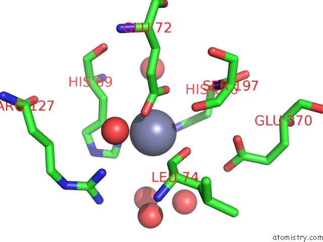
Mono view
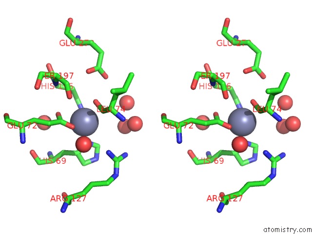
Stereo pair view
Mono view
Stereo pair view
A full contact list of Zinc with other atoms in the Zn binding
site number 1 of Crystal Structure of the Tick Carboxypeptidase Inhibitor in Complex with Human Carboxypeptidase B within 5.0Å range:
|
Reference:
J.L.Arolas,
G.M.Popowicz,
J.Lorenzo,
C.P.Sommerhoff,
R.Huber,
F.X.Aviles,
T.A.Holak.
The Three-Dimensional Structures of Tick Carboxypeptidase Inhibitor in Complex with A/B Carboxypeptidases Reveal A Novel Double-Headed Binding Mode J.Mol.Biol. V. 350 489 2005.
ISSN: ISSN 0022-2836
PubMed: 15961103
DOI: 10.1016/J.JMB.2005.05.015
Page generated: Wed Oct 16 21:18:59 2024
ISSN: ISSN 0022-2836
PubMed: 15961103
DOI: 10.1016/J.JMB.2005.05.015
Last articles
Zn in 9MJ5Zn in 9HNW
Zn in 9G0L
Zn in 9FNE
Zn in 9DZN
Zn in 9E0I
Zn in 9D32
Zn in 9DAK
Zn in 8ZXC
Zn in 8ZUF