Zinc »
PDB 1x8h-1xm8 »
1xa6 »
Zinc in PDB 1xa6: Crystal Structure of the Human BETA2-Chimaerin
Protein crystallography data
The structure of Crystal Structure of the Human BETA2-Chimaerin, PDB code: 1xa6
was solved by
B.Canagarajah,
F.C.Leskow,
J.Y.Ho,
H.Mischak,
L.F.Saidi,
M.G.Kazanietz,
J.H.Hurley,
with X-Ray Crystallography technique. A brief refinement statistics is given in the table below:
| Resolution Low / High (Å) | 20.00 / 3.20 |
| Space group | P 61 2 2 |
| Cell size a, b, c (Å), α, β, γ (°) | 131.090, 131.090, 288.750, 90.00, 90.00, 120.00 |
| R / Rfree (%) | 25.2 / 29.8 |
Zinc Binding Sites:
The binding sites of Zinc atom in the Crystal Structure of the Human BETA2-Chimaerin
(pdb code 1xa6). This binding sites where shown within
5.0 Angstroms radius around Zinc atom.
In total 2 binding sites of Zinc where determined in the Crystal Structure of the Human BETA2-Chimaerin, PDB code: 1xa6:
Jump to Zinc binding site number: 1; 2;
In total 2 binding sites of Zinc where determined in the Crystal Structure of the Human BETA2-Chimaerin, PDB code: 1xa6:
Jump to Zinc binding site number: 1; 2;
Zinc binding site 1 out of 2 in 1xa6
Go back to
Zinc binding site 1 out
of 2 in the Crystal Structure of the Human BETA2-Chimaerin
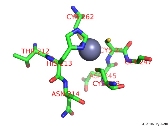
Mono view
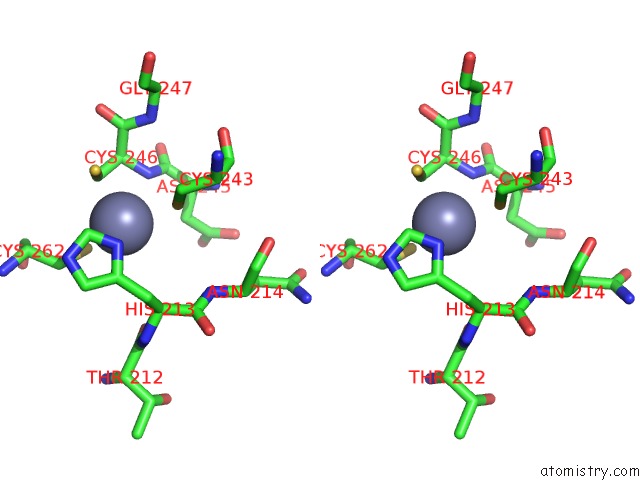
Stereo pair view
Mono view
Stereo pair view
|
|
A full contact list of Zinc with other atoms in the Zn binding
site number 1 of Crystal Structure of the Human BETA2-Chimaerin within 5.0Å range:
|
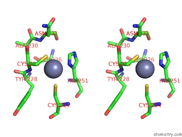
Zinc binding site 2 out of 2 in 1xa6
Go back to
Zinc binding site 2 out
of 2 in the Crystal Structure of the Human BETA2-Chimaerin
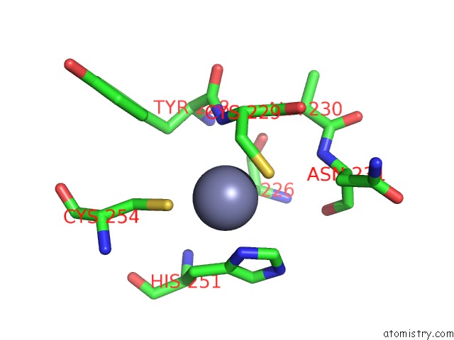
Mono view
Stereo pair view
Mono view
Stereo pair view
|
|
A full contact list of Zinc with other atoms in the Zn binding
site number 2 of Crystal Structure of the Human BETA2-Chimaerin within 5.0Å range:
|
Reference:
B.Canagarajah,
F.C.Leskow,
J.Y.Ho,
H.Mischak,
L.F.Saidi,
M.G.Kazanietz,
J.H.Hurley.
Structural Mechanism For Lipid Activation of the Rac-Specific Gap, BETA2-Chimaerin. Cell(Cambridge,Mass.) V. 119 407 2004.
ISSN: ISSN 0092-8674
PubMed: 15507211
DOI: 10.1016/J.CELL.2004.10.012
Page generated: Wed Oct 16 20:20:19 2024
ISSN: ISSN 0092-8674
PubMed: 15507211
DOI: 10.1016/J.CELL.2004.10.012
Last articles
Zn in 9MJ5Zn in 9HNW
Zn in 9G0L
Zn in 9FNE
Zn in 9DZN
Zn in 9E0I
Zn in 9D32
Zn in 9DAK
Zn in 8ZXC
Zn in 8ZUF