Zinc »
PDB 1lgd-1m2n »
1lu0 »
Zinc in PDB 1lu0: Atomic Resolution Structure of Squash Trypsin Inhibitor: Unexpected Metal Coordination
Protein crystallography data
The structure of Atomic Resolution Structure of Squash Trypsin Inhibitor: Unexpected Metal Coordination, PDB code: 1lu0
was solved by
R.Thaimattam,
E.Tykarska,
A.Bierzynski,
G.M.Sheldrick,
M.Jaskolski,
with X-Ray Crystallography technique. A brief refinement statistics is given in the table below:
| Resolution Low / High (Å) | 10.00 / 1.03 |
| Space group | P 21 21 2 |
| Cell size a, b, c (Å), α, β, γ (°) | 47.280, 58.630, 19.320, 90.00, 90.00, 90.00 |
| R / Rfree (%) | 12 / 14.5 |
Zinc Binding Sites:
The binding sites of Zinc atom in the Atomic Resolution Structure of Squash Trypsin Inhibitor: Unexpected Metal Coordination
(pdb code 1lu0). This binding sites where shown within
5.0 Angstroms radius around Zinc atom.
In total only one binding site of Zinc was determined in the Atomic Resolution Structure of Squash Trypsin Inhibitor: Unexpected Metal Coordination, PDB code: 1lu0:
In total only one binding site of Zinc was determined in the Atomic Resolution Structure of Squash Trypsin Inhibitor: Unexpected Metal Coordination, PDB code: 1lu0:
Zinc binding site 1 out of 1 in 1lu0
Go back to
Zinc binding site 1 out
of 1 in the Atomic Resolution Structure of Squash Trypsin Inhibitor: Unexpected Metal Coordination
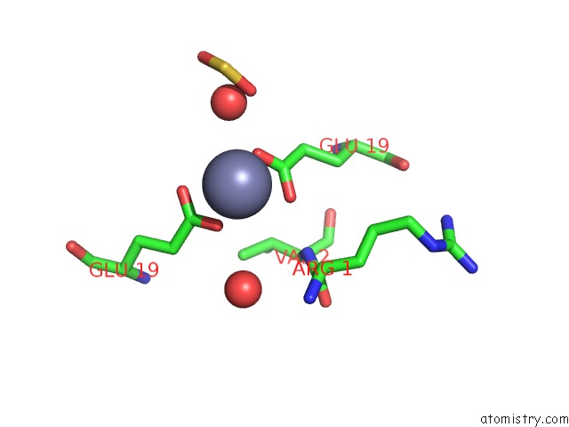
Mono view
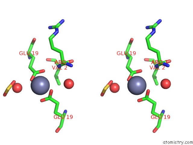
Stereo pair view
Mono view
Stereo pair view
A full contact list of Zinc with other atoms in the Zn binding
site number 1 of Atomic Resolution Structure of Squash Trypsin Inhibitor: Unexpected Metal Coordination within 5.0Å range:
|
Reference:
R.Thaimattam,
E.Tykarska,
A.Bierzynski,
G.M.Sheldrick,
M.Jaskolski.
Atomic Resolution Structure of Squash Trypsin Inhibitor: Unexpected Metal Coordination. Acta Crystallogr.,Sect.D V. 58 1448 2002.
ISSN: ISSN 0907-4449
PubMed: 12198301
DOI: 10.1107/S0907444902011769
Page generated: Sun Oct 13 05:14:00 2024
ISSN: ISSN 0907-4449
PubMed: 12198301
DOI: 10.1107/S0907444902011769
Last articles
Zn in 9MJ5Zn in 9HNW
Zn in 9G0L
Zn in 9FNE
Zn in 9DZN
Zn in 9E0I
Zn in 9D32
Zn in 9DAK
Zn in 8ZXC
Zn in 8ZUF