Zinc »
PDB 1lgd-1m2n »
1lru »
Zinc in PDB 1lru: Crystal Structure of E.Coli Peptide Deformylase Complexed with Antibiotic Actinonin
Enzymatic activity of Crystal Structure of E.Coli Peptide Deformylase Complexed with Antibiotic Actinonin
All present enzymatic activity of Crystal Structure of E.Coli Peptide Deformylase Complexed with Antibiotic Actinonin:
3.5.1.88;
3.5.1.88;
Protein crystallography data
The structure of Crystal Structure of E.Coli Peptide Deformylase Complexed with Antibiotic Actinonin, PDB code: 1lru
was solved by
J.-P.Guilloteau,
M.Mathieu,
C.Giglione,
V.Blanc,
A.Dupuy,
M.Chevrier,
P.Gil,
A.Famechon,
T.Meinnel,
V.Mikol,
with X-Ray Crystallography technique. A brief refinement statistics is given in the table below:
| Resolution Low / High (Å) | 15.00 / 2.10 |
| Space group | C 1 2 1 |
| Cell size a, b, c (Å), α, β, γ (°) | 141.040, 63.608, 83.494, 90.00, 123.35, 90.00 |
| R / Rfree (%) | 24 / n/a |
Zinc Binding Sites:
The binding sites of Zinc atom in the Crystal Structure of E.Coli Peptide Deformylase Complexed with Antibiotic Actinonin
(pdb code 1lru). This binding sites where shown within
5.0 Angstroms radius around Zinc atom.
In total 3 binding sites of Zinc where determined in the Crystal Structure of E.Coli Peptide Deformylase Complexed with Antibiotic Actinonin, PDB code: 1lru:
Jump to Zinc binding site number: 1; 2; 3;
In total 3 binding sites of Zinc where determined in the Crystal Structure of E.Coli Peptide Deformylase Complexed with Antibiotic Actinonin, PDB code: 1lru:
Jump to Zinc binding site number: 1; 2; 3;
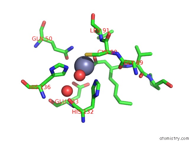
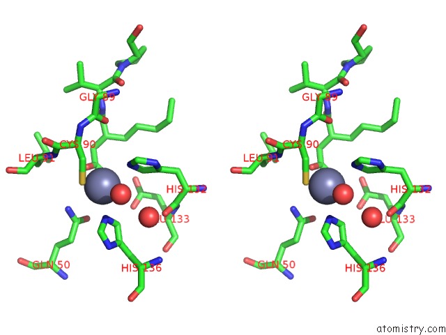
Zinc binding site 1 out of 3 in 1lru
Go back to
Zinc binding site 1 out
of 3 in the Crystal Structure of E.Coli Peptide Deformylase Complexed with Antibiotic Actinonin
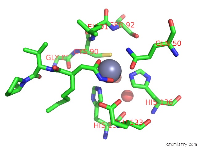
Mono view
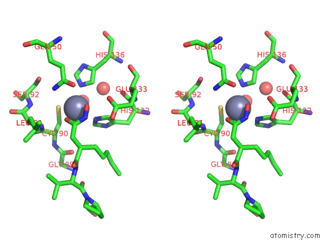
Stereo pair view
Mono view
Stereo pair view
A full contact list of Zinc with other atoms in the Zn binding
site number 1 of Crystal Structure of E.Coli Peptide Deformylase Complexed with Antibiotic Actinonin within 5.0Å range:
|
Zinc binding site 2 out of 3 in 1lru
Go back to
Zinc binding site 2 out
of 3 in the Crystal Structure of E.Coli Peptide Deformylase Complexed with Antibiotic Actinonin
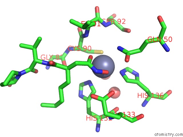
Mono view
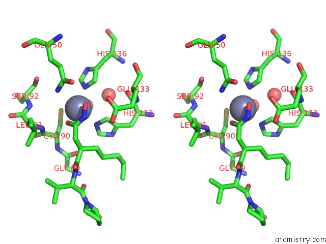
Stereo pair view
Mono view
Stereo pair view
A full contact list of Zinc with other atoms in the Zn binding
site number 2 of Crystal Structure of E.Coli Peptide Deformylase Complexed with Antibiotic Actinonin within 5.0Å range:
|
Zinc binding site 3 out of 3 in 1lru
Go back to
Zinc binding site 3 out
of 3 in the Crystal Structure of E.Coli Peptide Deformylase Complexed with Antibiotic Actinonin
Mono view
Stereo pair view
Mono view
Stereo pair view
A full contact list of Zinc with other atoms in the Zn binding
site number 3 of Crystal Structure of E.Coli Peptide Deformylase Complexed with Antibiotic Actinonin within 5.0Å range:
|
Reference:
J.P.Guilloteau,
M.Mathieu,
C.Giglione,
V.Blanc,
A.Dupuy,
M.Chevrier,
P.Gil,
A.Famechon,
T.Meinnel,
V.Mikol.
The Crystal Structures of Four Peptide Deformylases Bound to the Antibiotic Actinonin Reveal Two Distinct Types: A Platform For the Structure-Based Design of Antibacterial Agents. J.Mol.Biol. V. 320 951 2002.
ISSN: ISSN 0022-2836
PubMed: 12126617
DOI: 10.1016/S0022-2836(02)00549-1
Page generated: Sun Oct 13 05:10:52 2024
ISSN: ISSN 0022-2836
PubMed: 12126617
DOI: 10.1016/S0022-2836(02)00549-1
Last articles
Zn in 9MJ5Zn in 9HNW
Zn in 9G0L
Zn in 9FNE
Zn in 9DZN
Zn in 9E0I
Zn in 9D32
Zn in 9DAK
Zn in 8ZXC
Zn in 8ZUF