Zinc »
PDB 1kk1-1kzp »
1kol »
Zinc in PDB 1kol: Crystal Structure of Formaldehyde Dehydrogenase
Enzymatic activity of Crystal Structure of Formaldehyde Dehydrogenase
All present enzymatic activity of Crystal Structure of Formaldehyde Dehydrogenase:
1.2.1.46;
1.2.1.46;
Protein crystallography data
The structure of Crystal Structure of Formaldehyde Dehydrogenase, PDB code: 1kol
was solved by
N.Tanaka,
Y.Kusakabe,
K.Ito,
T.Yoshimoto,
K.T.Nakamura,
with X-Ray Crystallography technique. A brief refinement statistics is given in the table below:
| Resolution Low / High (Å) | 30.00 / 1.65 |
| Space group | P 31 1 2 |
| Cell size a, b, c (Å), α, β, γ (°) | 85.686, 85.686, 190.752, 90.00, 90.00, 120.00 |
| R / Rfree (%) | 17.1 / 20.6 |
Zinc Binding Sites:
The binding sites of Zinc atom in the Crystal Structure of Formaldehyde Dehydrogenase
(pdb code 1kol). This binding sites where shown within
5.0 Angstroms radius around Zinc atom.
In total 4 binding sites of Zinc where determined in the Crystal Structure of Formaldehyde Dehydrogenase, PDB code: 1kol:
Jump to Zinc binding site number: 1; 2; 3; 4;
In total 4 binding sites of Zinc where determined in the Crystal Structure of Formaldehyde Dehydrogenase, PDB code: 1kol:
Jump to Zinc binding site number: 1; 2; 3; 4;
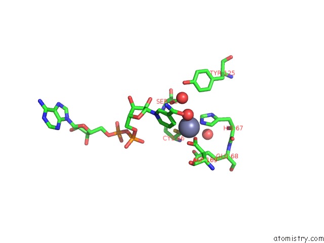
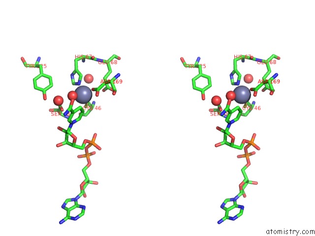
Zinc binding site 1 out of 4 in 1kol
Go back to
Zinc binding site 1 out
of 4 in the Crystal Structure of Formaldehyde Dehydrogenase
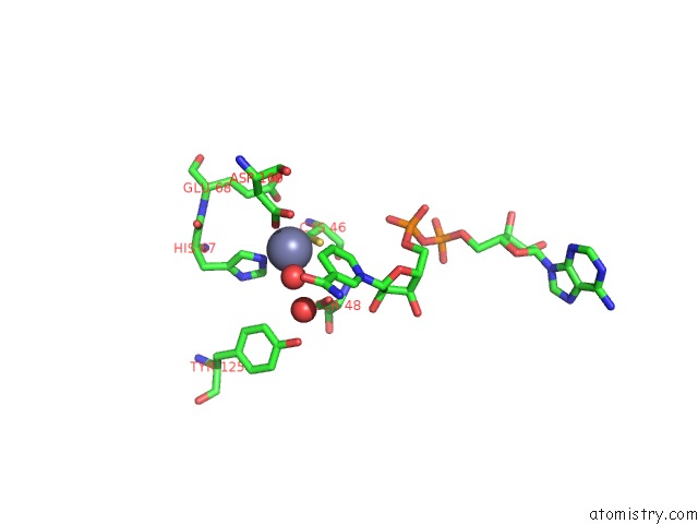
Mono view
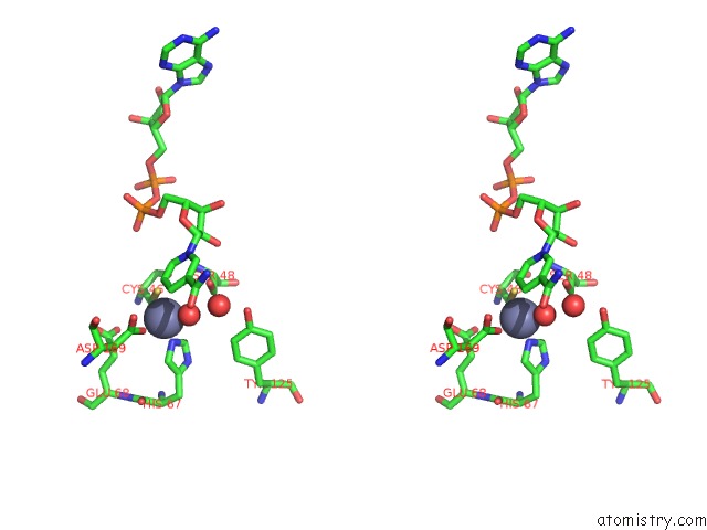
Stereo pair view
Mono view
Stereo pair view
A full contact list of Zinc with other atoms in the Zn binding
site number 1 of Crystal Structure of Formaldehyde Dehydrogenase within 5.0Å range:
|
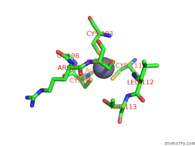
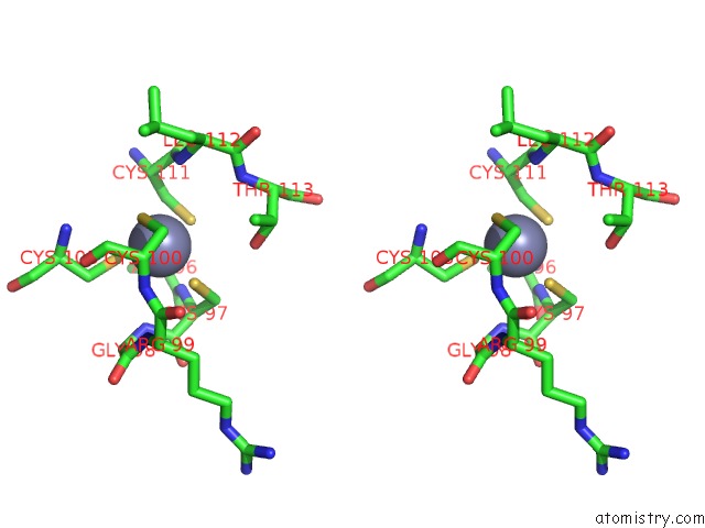
Zinc binding site 2 out of 4 in 1kol
Go back to
Zinc binding site 2 out
of 4 in the Crystal Structure of Formaldehyde Dehydrogenase
Mono view
Stereo pair view
Mono view
Stereo pair view
A full contact list of Zinc with other atoms in the Zn binding
site number 2 of Crystal Structure of Formaldehyde Dehydrogenase within 5.0Å range:
|
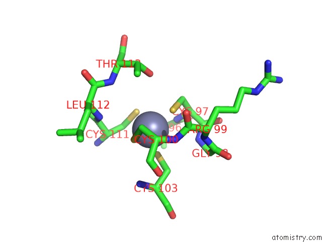
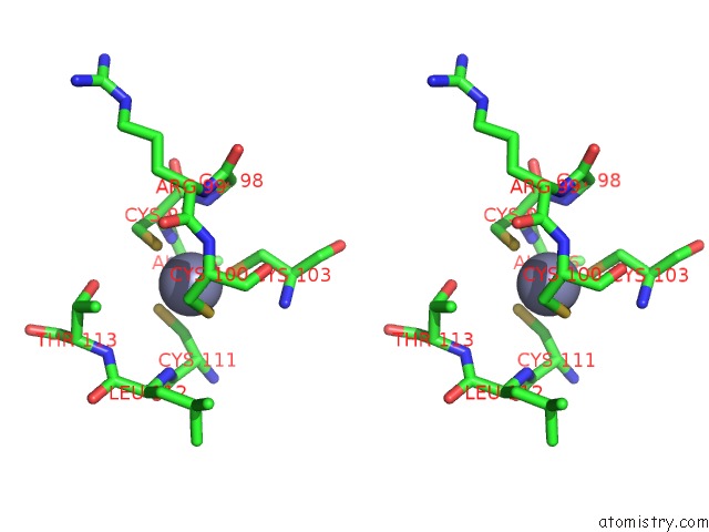
Zinc binding site 3 out of 4 in 1kol
Go back to
Zinc binding site 3 out
of 4 in the Crystal Structure of Formaldehyde Dehydrogenase
Mono view
Stereo pair view
Mono view
Stereo pair view
A full contact list of Zinc with other atoms in the Zn binding
site number 3 of Crystal Structure of Formaldehyde Dehydrogenase within 5.0Å range:
|
Zinc binding site 4 out of 4 in 1kol
Go back to
Zinc binding site 4 out
of 4 in the Crystal Structure of Formaldehyde Dehydrogenase
Mono view
Stereo pair view
Mono view
Stereo pair view
A full contact list of Zinc with other atoms in the Zn binding
site number 4 of Crystal Structure of Formaldehyde Dehydrogenase within 5.0Å range:
|
Reference:
N.Tanaka,
Y.Kusakabe,
K.Ito,
T.Yoshimoto,
K.T.Nakamura.
Crystal Structure of Formaldehyde Dehydrogenase From Pseudomonas Putida: the Structural Origin of the Tightly Bound Cofactor in Nicotinoprotein Dehydrogenases J.Mol.Biol. V. 324 519 2002.
ISSN: ISSN 0022-2836
PubMed: 12445786
DOI: 10.1016/S0022-2836(02)01066-5
Page generated: Sun Oct 13 04:35:29 2024
ISSN: ISSN 0022-2836
PubMed: 12445786
DOI: 10.1016/S0022-2836(02)01066-5
Last articles
Zn in 9MJ5Zn in 9HNW
Zn in 9G0L
Zn in 9FNE
Zn in 9DZN
Zn in 9E0I
Zn in 9D32
Zn in 9DAK
Zn in 8ZXC
Zn in 8ZUF