Zinc »
PDB 1k6y-1kk0 »
1k82 »
Zinc in PDB 1k82: Crystal Structure of E.Coli Formamidopyrimidine-Dna Glycosylase (Fpg) Covalently Trapped with Dna
Enzymatic activity of Crystal Structure of E.Coli Formamidopyrimidine-Dna Glycosylase (Fpg) Covalently Trapped with Dna
All present enzymatic activity of Crystal Structure of E.Coli Formamidopyrimidine-Dna Glycosylase (Fpg) Covalently Trapped with Dna:
3.2.2.23;
3.2.2.23;
Protein crystallography data
The structure of Crystal Structure of E.Coli Formamidopyrimidine-Dna Glycosylase (Fpg) Covalently Trapped with Dna, PDB code: 1k82
was solved by
R.Gilboa,
D.O.Zharkov,
G.Golan,
A.S.Fernandes,
S.E.Gerchman,
E.Matz,
J.H.Kycia,
A.P.Grollman,
G.Shoham,
with X-Ray Crystallography technique. A brief refinement statistics is given in the table below:
| Resolution Low / High (Å) | 34.00 / 2.10 |
| Space group | P 1 21 1 |
| Cell size a, b, c (Å), α, β, γ (°) | 80.704, 96.033, 96.228, 90.00, 96.80, 90.00 |
| R / Rfree (%) | 21.4 / 26.5 |
Zinc Binding Sites:
The binding sites of Zinc atom in the Crystal Structure of E.Coli Formamidopyrimidine-Dna Glycosylase (Fpg) Covalently Trapped with Dna
(pdb code 1k82). This binding sites where shown within
5.0 Angstroms radius around Zinc atom.
In total 4 binding sites of Zinc where determined in the Crystal Structure of E.Coli Formamidopyrimidine-Dna Glycosylase (Fpg) Covalently Trapped with Dna, PDB code: 1k82:
Jump to Zinc binding site number: 1; 2; 3; 4;
In total 4 binding sites of Zinc where determined in the Crystal Structure of E.Coli Formamidopyrimidine-Dna Glycosylase (Fpg) Covalently Trapped with Dna, PDB code: 1k82:
Jump to Zinc binding site number: 1; 2; 3; 4;
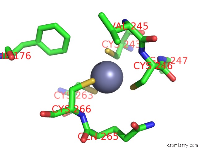
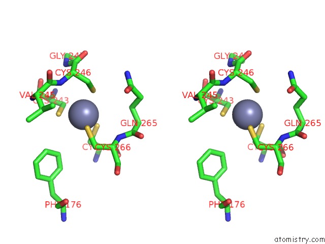
Zinc binding site 1 out of 4 in 1k82
Go back to
Zinc binding site 1 out
of 4 in the Crystal Structure of E.Coli Formamidopyrimidine-Dna Glycosylase (Fpg) Covalently Trapped with Dna

Mono view

Stereo pair view
Mono view
Stereo pair view
A full contact list of Zinc with other atoms in the Zn binding
site number 1 of Crystal Structure of E.Coli Formamidopyrimidine-Dna Glycosylase (Fpg) Covalently Trapped with Dna within 5.0Å range:
|
Zinc binding site 2 out of 4 in 1k82
Go back to
Zinc binding site 2 out
of 4 in the Crystal Structure of E.Coli Formamidopyrimidine-Dna Glycosylase (Fpg) Covalently Trapped with Dna

Mono view

Stereo pair view
Mono view
Stereo pair view
A full contact list of Zinc with other atoms in the Zn binding
site number 2 of Crystal Structure of E.Coli Formamidopyrimidine-Dna Glycosylase (Fpg) Covalently Trapped with Dna within 5.0Å range:
|
Zinc binding site 3 out of 4 in 1k82
Go back to
Zinc binding site 3 out
of 4 in the Crystal Structure of E.Coli Formamidopyrimidine-Dna Glycosylase (Fpg) Covalently Trapped with Dna
Mono view
Stereo pair view
Mono view
Stereo pair view
A full contact list of Zinc with other atoms in the Zn binding
site number 3 of Crystal Structure of E.Coli Formamidopyrimidine-Dna Glycosylase (Fpg) Covalently Trapped with Dna within 5.0Å range:
|
Zinc binding site 4 out of 4 in 1k82
Go back to
Zinc binding site 4 out
of 4 in the Crystal Structure of E.Coli Formamidopyrimidine-Dna Glycosylase (Fpg) Covalently Trapped with Dna

Mono view

Stereo pair view
Mono view
Stereo pair view
A full contact list of Zinc with other atoms in the Zn binding
site number 4 of Crystal Structure of E.Coli Formamidopyrimidine-Dna Glycosylase (Fpg) Covalently Trapped with Dna within 5.0Å range:
|
Reference:
R.Gilboa,
D.O.Zharkov,
G.Golan,
A.S.Fernandes,
S.E.Gerchman,
E.Matz,
J.H.Kycia,
A.P.Grollman,
G.Shoham.
Structure of Formamidopyrimidine-Dna Glycosylase Covalently Complexed to Dna. J.Biol.Chem. V. 277 19811 2002.
ISSN: ISSN 0021-9258
PubMed: 11912217
DOI: 10.1074/JBC.M202058200
Page generated: Sun Oct 13 04:09:13 2024
ISSN: ISSN 0021-9258
PubMed: 11912217
DOI: 10.1074/JBC.M202058200
Last articles
Zn in 9MJ5Zn in 9HNW
Zn in 9G0L
Zn in 9FNE
Zn in 9DZN
Zn in 9E0I
Zn in 9D32
Zn in 9DAK
Zn in 8ZXC
Zn in 8ZUF