Zinc »
PDB 1jpu-1k53 »
1k2t »
Zinc in PDB 1k2t: Structure of Rat Brain Nnos Heme Domain Complexed with S- Ethyl-N-Phenyl-Isothiourea
Enzymatic activity of Structure of Rat Brain Nnos Heme Domain Complexed with S- Ethyl-N-Phenyl-Isothiourea
All present enzymatic activity of Structure of Rat Brain Nnos Heme Domain Complexed with S- Ethyl-N-Phenyl-Isothiourea:
1.14.13.39;
1.14.13.39;
Protein crystallography data
The structure of Structure of Rat Brain Nnos Heme Domain Complexed with S- Ethyl-N-Phenyl-Isothiourea, PDB code: 1k2t
was solved by
H.Li,
P.Martasek,
B.S.S.Masters,
T.L.Poulos,
C.S.Raman,
with X-Ray Crystallography technique. A brief refinement statistics is given in the table below:
| Resolution Low / High (Å) | 29.70 / 2.20 |
| Space group | P 21 21 21 |
| Cell size a, b, c (Å), α, β, γ (°) | 52.000, 111.240, 165.000, 90.00, 90.00, 90.00 |
| R / Rfree (%) | 22.4 / 26.5 |
Other elements in 1k2t:
The structure of Structure of Rat Brain Nnos Heme Domain Complexed with S- Ethyl-N-Phenyl-Isothiourea also contains other interesting chemical elements:
| Iron | (Fe) | 2 atoms |
Zinc Binding Sites:
The binding sites of Zinc atom in the Structure of Rat Brain Nnos Heme Domain Complexed with S- Ethyl-N-Phenyl-Isothiourea
(pdb code 1k2t). This binding sites where shown within
5.0 Angstroms radius around Zinc atom.
In total only one binding site of Zinc was determined in the Structure of Rat Brain Nnos Heme Domain Complexed with S- Ethyl-N-Phenyl-Isothiourea, PDB code: 1k2t:
In total only one binding site of Zinc was determined in the Structure of Rat Brain Nnos Heme Domain Complexed with S- Ethyl-N-Phenyl-Isothiourea, PDB code: 1k2t:
Zinc binding site 1 out of 1 in 1k2t
Go back to
Zinc binding site 1 out
of 1 in the Structure of Rat Brain Nnos Heme Domain Complexed with S- Ethyl-N-Phenyl-Isothiourea
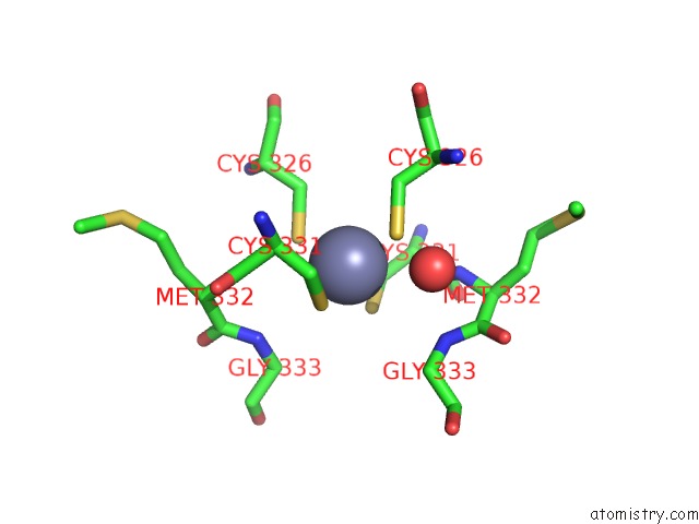
Mono view
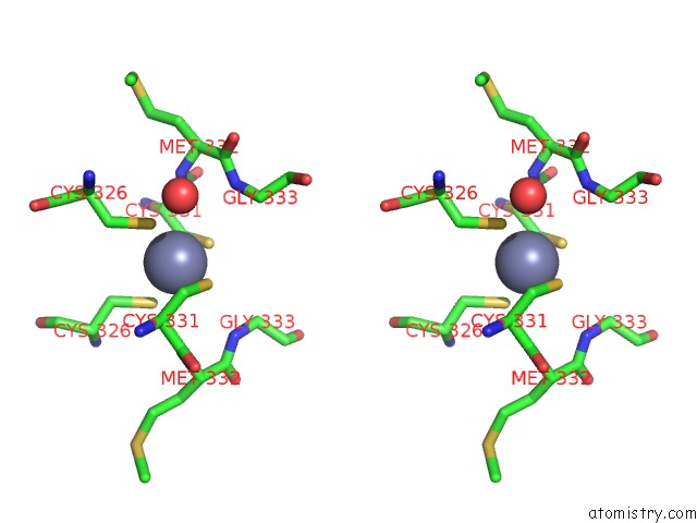
Stereo pair view
Mono view
Stereo pair view
A full contact list of Zinc with other atoms in the Zn binding
site number 1 of Structure of Rat Brain Nnos Heme Domain Complexed with S- Ethyl-N-Phenyl-Isothiourea within 5.0Å range:
|
Reference:
H.Li,
P.Martasek,
B.S.S.Masters,
T.L.Poulos,
C.S.Raman.
Structure of Rat Brain Nnos Heme Domain To Be Published.
Page generated: Tue Aug 19 21:11:43 2025
Last articles
Zn in 1X4LZn in 1X4K
Zn in 1X4J
Zn in 1X4I
Zn in 1X3H
Zn in 1X3Z
Zn in 1X3W
Zn in 1X3C
Zn in 1X1C
Zn in 1X1V