Zinc »
PDB 1wwe-1x8g »
1x1c »
Zinc in PDB 1x1c: Crystal Structure of Bchu Complexed with S-Adenosyl-L-Homocysteine and ZN2+
Protein crystallography data
The structure of Crystal Structure of Bchu Complexed with S-Adenosyl-L-Homocysteine and ZN2+, PDB code: 1x1c
was solved by
H.Yamaguchi,
K.Wada,
K.Fukuyama,
with X-Ray Crystallography technique. A brief refinement statistics is given in the table below:
| Resolution Low / High (Å) | 41.83 / 2.85 |
| Space group | P 65 2 2 |
| Cell size a, b, c (Å), α, β, γ (°) | 81.615, 81.615, 250.960, 90.00, 90.00, 120.00 |
| R / Rfree (%) | 23 / 28.8 |
Zinc Binding Sites:
The binding sites of Zinc atom in the Crystal Structure of Bchu Complexed with S-Adenosyl-L-Homocysteine and ZN2+
(pdb code 1x1c). This binding sites where shown within
5.0 Angstroms radius around Zinc atom.
In total 5 binding sites of Zinc where determined in the Crystal Structure of Bchu Complexed with S-Adenosyl-L-Homocysteine and ZN2+, PDB code: 1x1c:
Jump to Zinc binding site number: 1; 2; 3; 4; 5;
In total 5 binding sites of Zinc where determined in the Crystal Structure of Bchu Complexed with S-Adenosyl-L-Homocysteine and ZN2+, PDB code: 1x1c:
Jump to Zinc binding site number: 1; 2; 3; 4; 5;
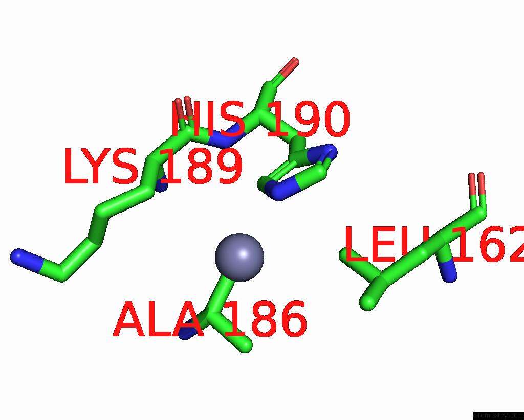
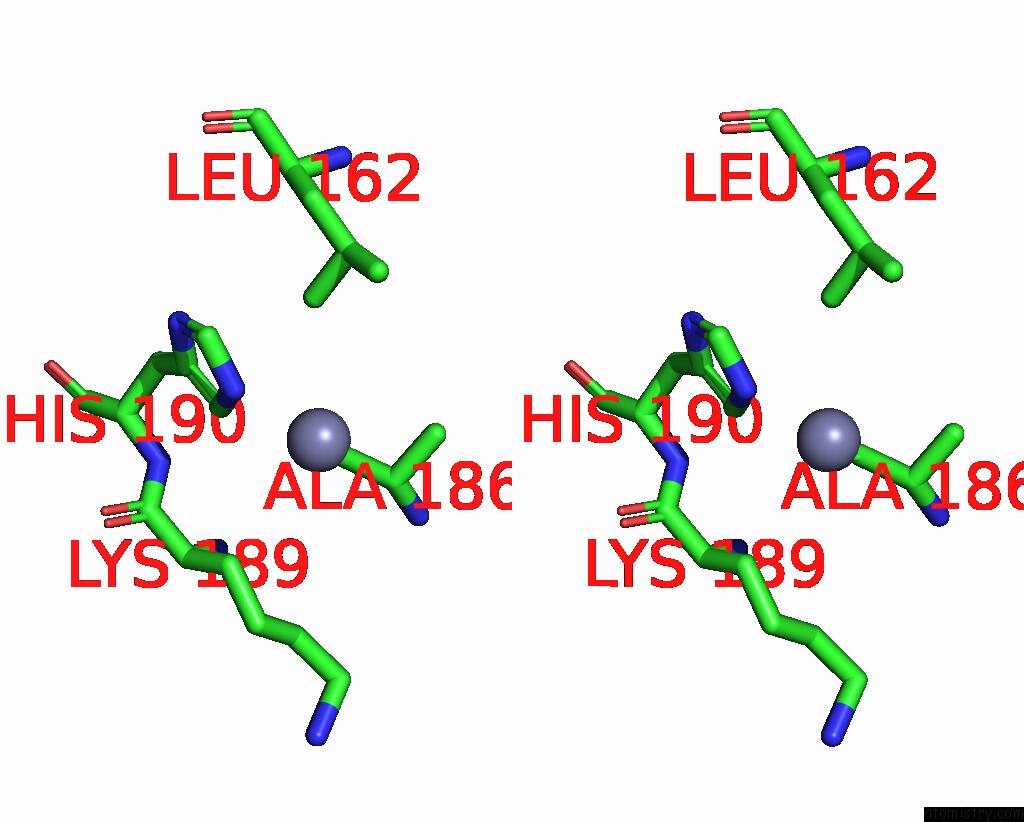
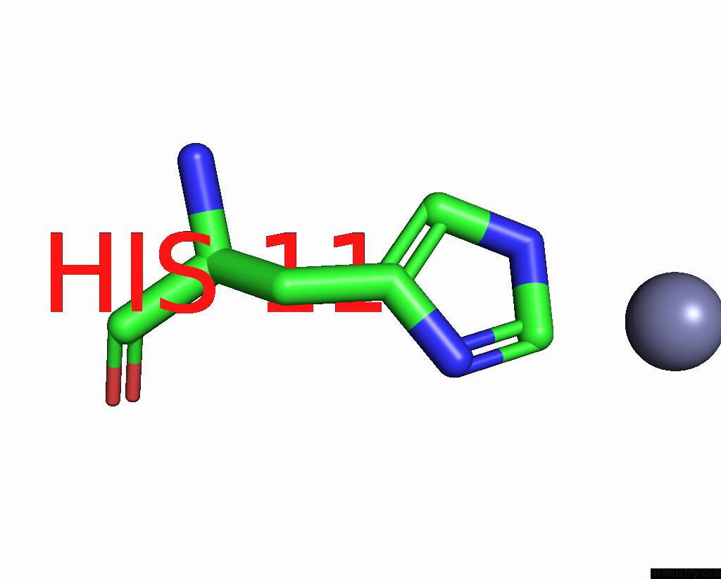
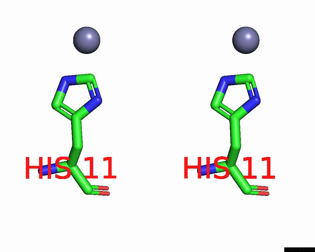
Zinc binding site 1 out of 5 in 1x1c
Go back to
Zinc binding site 1 out
of 5 in the Crystal Structure of Bchu Complexed with S-Adenosyl-L-Homocysteine and ZN2+
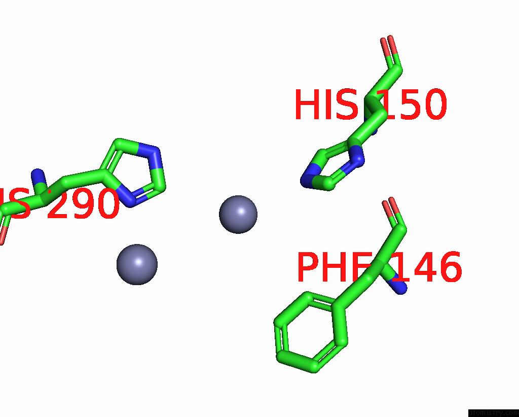
Mono view
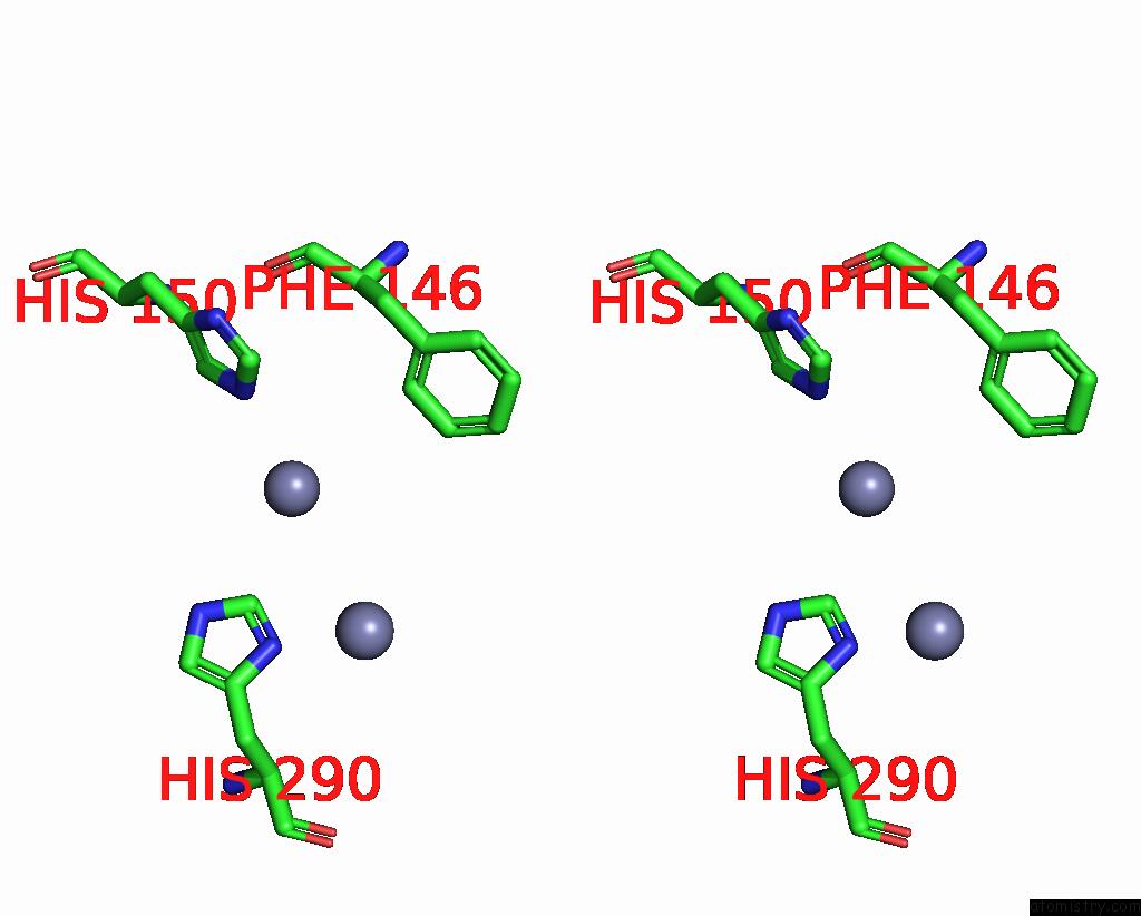
Stereo pair view
Mono view
Stereo pair view
A full contact list of Zinc with other atoms in the Zn binding
site number 1 of Crystal Structure of Bchu Complexed with S-Adenosyl-L-Homocysteine and ZN2+ within 5.0Å range:
|
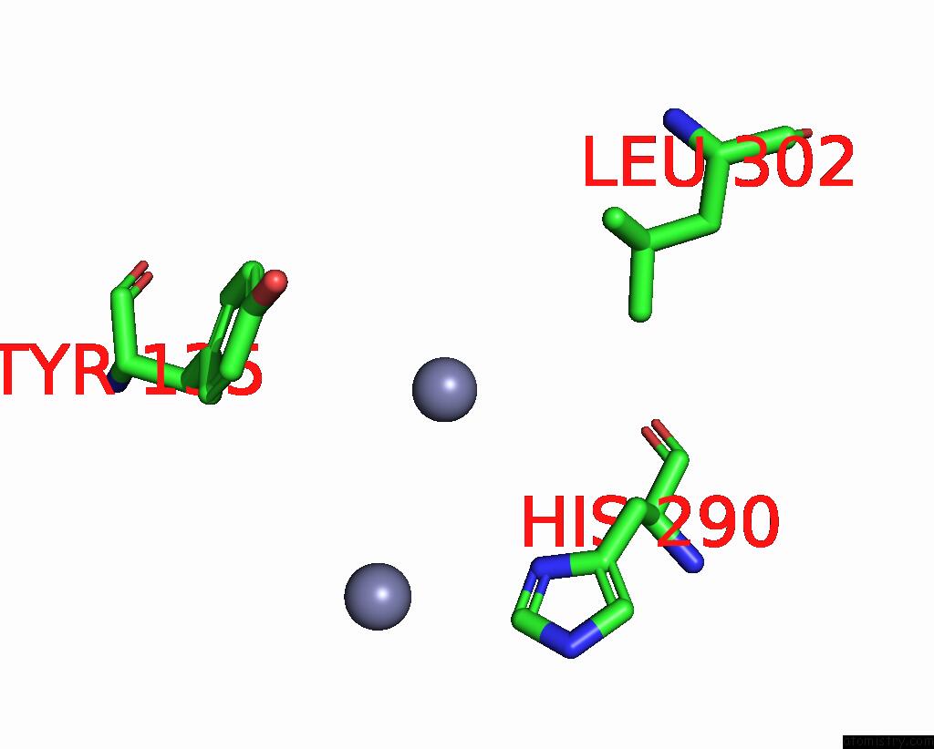
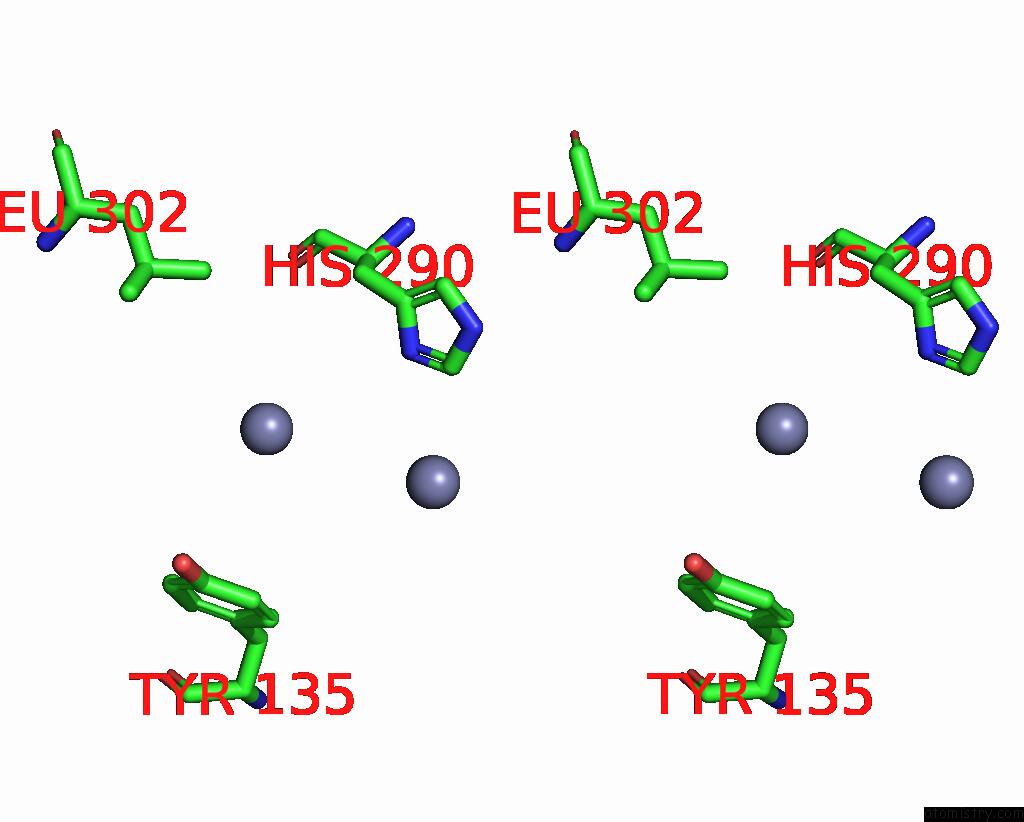
Zinc binding site 2 out of 5 in 1x1c
Go back to
Zinc binding site 2 out
of 5 in the Crystal Structure of Bchu Complexed with S-Adenosyl-L-Homocysteine and ZN2+
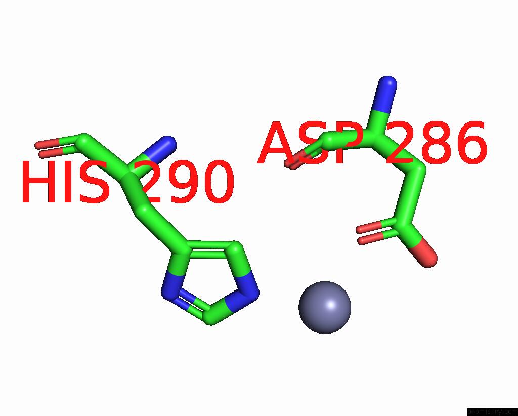
Mono view
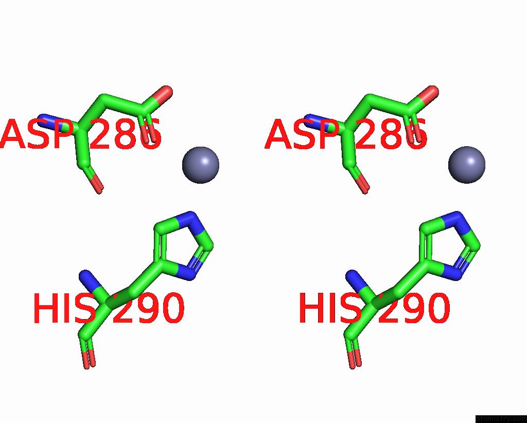
Stereo pair view
Mono view
Stereo pair view
A full contact list of Zinc with other atoms in the Zn binding
site number 2 of Crystal Structure of Bchu Complexed with S-Adenosyl-L-Homocysteine and ZN2+ within 5.0Å range:
|
Zinc binding site 3 out of 5 in 1x1c
Go back to
Zinc binding site 3 out
of 5 in the Crystal Structure of Bchu Complexed with S-Adenosyl-L-Homocysteine and ZN2+
Mono view
Stereo pair view
Mono view
Stereo pair view
A full contact list of Zinc with other atoms in the Zn binding
site number 3 of Crystal Structure of Bchu Complexed with S-Adenosyl-L-Homocysteine and ZN2+ within 5.0Å range:
|
Zinc binding site 4 out of 5 in 1x1c
Go back to
Zinc binding site 4 out
of 5 in the Crystal Structure of Bchu Complexed with S-Adenosyl-L-Homocysteine and ZN2+
Mono view
Stereo pair view
Mono view
Stereo pair view
A full contact list of Zinc with other atoms in the Zn binding
site number 4 of Crystal Structure of Bchu Complexed with S-Adenosyl-L-Homocysteine and ZN2+ within 5.0Å range:
|
Zinc binding site 5 out of 5 in 1x1c
Go back to
Zinc binding site 5 out
of 5 in the Crystal Structure of Bchu Complexed with S-Adenosyl-L-Homocysteine and ZN2+
Mono view
Stereo pair view
Mono view
Stereo pair view
A full contact list of Zinc with other atoms in the Zn binding
site number 5 of Crystal Structure of Bchu Complexed with S-Adenosyl-L-Homocysteine and ZN2+ within 5.0Å range:
|
Reference:
K.Wada,
H.Yamaguchi,
J.Harada,
K.Niimi,
S.Osumi,
Y.Saga,
H.Oh-Oka,
H.Tamiaki,
K.Fukuyama.
Crystal Structures of Bchu, A Methyltransferase Involved in Bacteriochlorophyll C Biosynthesis, and Its Complex with S-Adenosylhomocysteine: Implications For Reaction Mechanism. J.Mol.Biol. V. 360 839 2006.
ISSN: ISSN 0022-2836
PubMed: 16797589
DOI: 10.1016/J.JMB.2006.05.057
Page generated: Wed Aug 20 00:09:04 2025
ISSN: ISSN 0022-2836
PubMed: 16797589
DOI: 10.1016/J.JMB.2006.05.057
Last articles
Zn in 2G9TZn in 2GDA
Zn in 2GC3
Zn in 2GD8
Zn in 2GC2
Zn in 2GC1
Zn in 2GC0
Zn in 2GBX
Zn in 2GBV
Zn in 2GBT