Zinc »
PDB 1hxy-1i95 »
1i7w »
Zinc in PDB 1i7w: Beta-Catenin/Phosphorylated E-Cadherin Complex
Protein crystallography data
The structure of Beta-Catenin/Phosphorylated E-Cadherin Complex, PDB code: 1i7w
was solved by
A.H.Huber,
W.I.Weis,
with X-Ray Crystallography technique. A brief refinement statistics is given in the table below:
| Resolution Low / High (Å) | 100.00 / 2.00 |
| Space group | C 1 2 1 |
| Cell size a, b, c (Å), α, β, γ (°) | 168.600, 85.300, 115.100, 90.00, 107.90, 90.00 |
| R / Rfree (%) | 20.6 / 24.5 |
Other elements in 1i7w:
The structure of Beta-Catenin/Phosphorylated E-Cadherin Complex also contains other interesting chemical elements:
| Chlorine | (Cl) | 8 atoms |
Zinc Binding Sites:
The binding sites of Zinc atom in the Beta-Catenin/Phosphorylated E-Cadherin Complex
(pdb code 1i7w). This binding sites where shown within
5.0 Angstroms radius around Zinc atom.
In total 2 binding sites of Zinc where determined in the Beta-Catenin/Phosphorylated E-Cadherin Complex, PDB code: 1i7w:
Jump to Zinc binding site number: 1; 2;
In total 2 binding sites of Zinc where determined in the Beta-Catenin/Phosphorylated E-Cadherin Complex, PDB code: 1i7w:
Jump to Zinc binding site number: 1; 2;
Zinc binding site 1 out of 2 in 1i7w
Go back to
Zinc binding site 1 out
of 2 in the Beta-Catenin/Phosphorylated E-Cadherin Complex
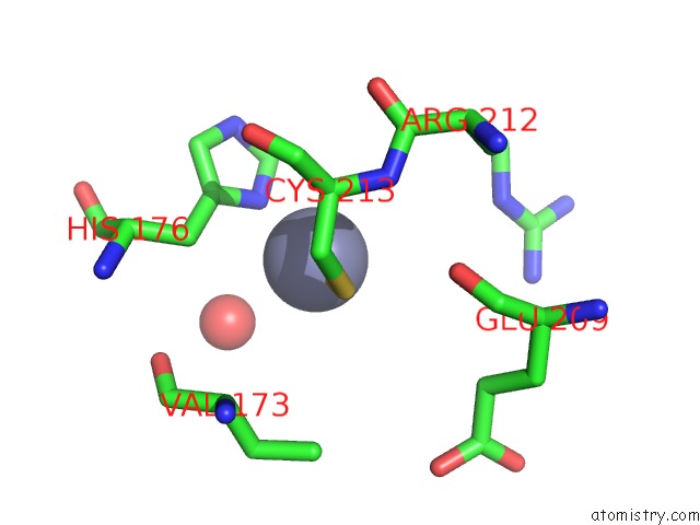
Mono view
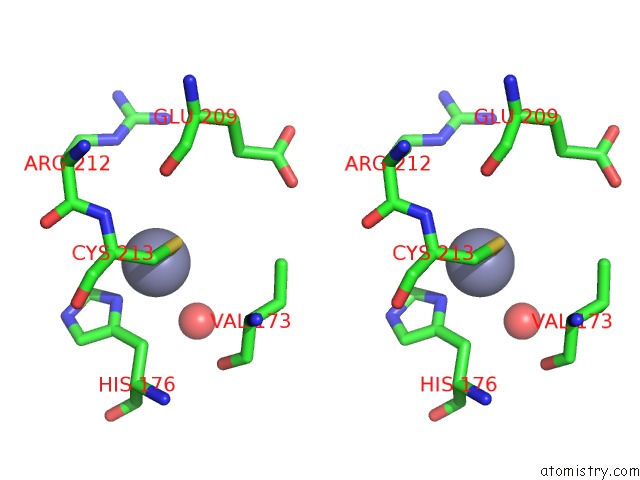
Stereo pair view
Mono view
Stereo pair view
A full contact list of Zinc with other atoms in the Zn binding
site number 1 of Beta-Catenin/Phosphorylated E-Cadherin Complex within 5.0Å range:
|
Zinc binding site 2 out of 2 in 1i7w
Go back to
Zinc binding site 2 out
of 2 in the Beta-Catenin/Phosphorylated E-Cadherin Complex
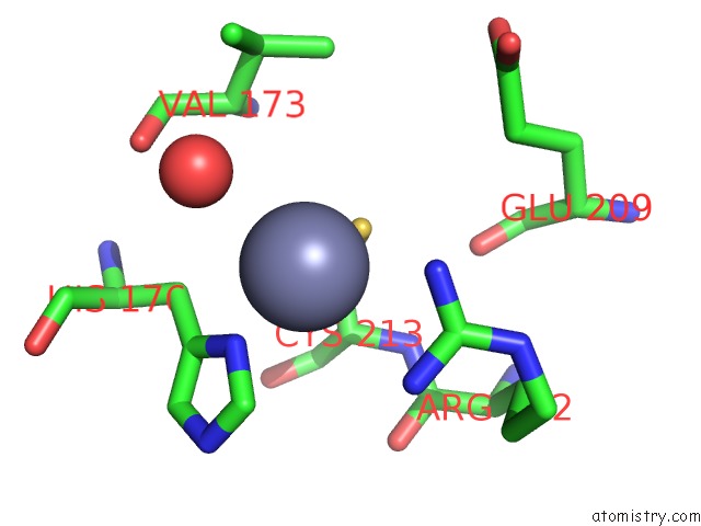
Mono view
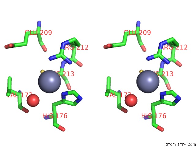
Stereo pair view
Mono view
Stereo pair view
A full contact list of Zinc with other atoms in the Zn binding
site number 2 of Beta-Catenin/Phosphorylated E-Cadherin Complex within 5.0Å range:
|
Reference:
A.H.Huber,
W.I.Weis.
The Structure of the Beta-Catenin/E-Cadherin Complex and the Molecular Basis of Diverse Ligand Recognition By Beta-Catenin. Cell(Cambridge,Mass.) V. 105 391 2001.
ISSN: ISSN 0092-8674
PubMed: 11348595
DOI: 10.1016/S0092-8674(01)00330-0
Page generated: Sun Oct 13 02:56:24 2024
ISSN: ISSN 0092-8674
PubMed: 11348595
DOI: 10.1016/S0092-8674(01)00330-0
Last articles
Zn in 9MJ5Zn in 9HNW
Zn in 9G0L
Zn in 9FNE
Zn in 9DZN
Zn in 9E0I
Zn in 9D32
Zn in 9DAK
Zn in 8ZXC
Zn in 8ZUF