Zinc »
PDB 3elc-3eyw »
3exl »
Zinc in PDB 3exl: Crystal Structure of A P53 Core Tetramer Bound to Dna
Protein crystallography data
The structure of Crystal Structure of A P53 Core Tetramer Bound to Dna, PDB code: 3exl
was solved by
K.A.Malecka,
with X-Ray Crystallography technique. A brief refinement statistics is given in the table below:
| Resolution Low / High (Å) | 57.35 / 2.20 |
| Space group | C 1 2 1 |
| Cell size a, b, c (Å), α, β, γ (°) | 109.418, 68.103, 34.428, 90.00, 104.18, 90.00 |
| R / Rfree (%) | 20.2 / 25.3 |
Zinc Binding Sites:
The binding sites of Zinc atom in the Crystal Structure of A P53 Core Tetramer Bound to Dna
(pdb code 3exl). This binding sites where shown within
5.0 Angstroms radius around Zinc atom.
In total only one binding site of Zinc was determined in the Crystal Structure of A P53 Core Tetramer Bound to Dna, PDB code: 3exl:
In total only one binding site of Zinc was determined in the Crystal Structure of A P53 Core Tetramer Bound to Dna, PDB code: 3exl:
Zinc binding site 1 out of 1 in 3exl
Go back to
Zinc binding site 1 out
of 1 in the Crystal Structure of A P53 Core Tetramer Bound to Dna
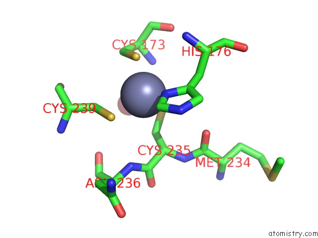
Mono view
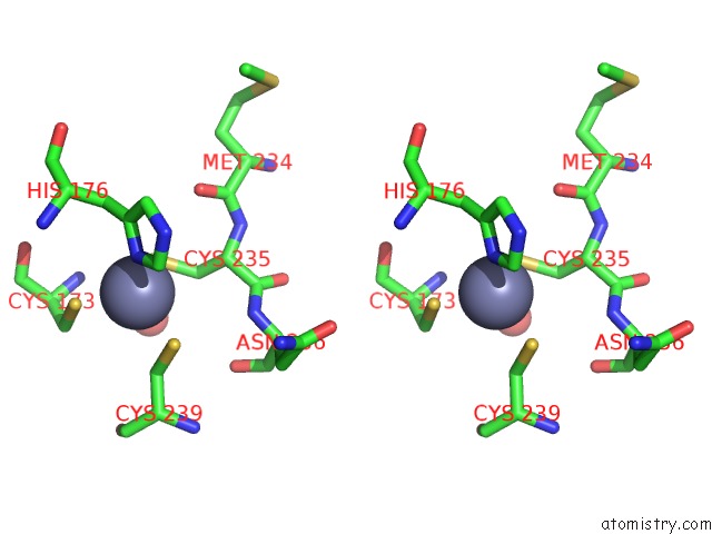
Stereo pair view
Mono view
Stereo pair view
A full contact list of Zinc with other atoms in the Zn binding
site number 1 of Crystal Structure of A P53 Core Tetramer Bound to Dna within 5.0Å range:
|
Reference:
K.A.Malecka,
W.C.Ho,
R.Marmorstein.
Crystal Structure of A P53 Core Tetramer Bound to Dna. Oncogene V. 28 325 2009.
ISSN: ISSN 0950-9232
PubMed: 18978813
DOI: 10.1038/ONC.2008.400
Page generated: Wed Aug 20 09:01:22 2025
ISSN: ISSN 0950-9232
PubMed: 18978813
DOI: 10.1038/ONC.2008.400
Last articles
Zn in 4B86Zn in 4B6D
Zn in 4BBP
Zn in 4BAC
Zn in 4B92
Zn in 4B9P
Zn in 4B87
Zn in 4B7Y
Zn in 4B56
Zn in 4B6Z