Zinc »
PDB 4b52-4bj9 »
4b86 »
Zinc in PDB 4b86: Crystal Structure of the MSL1-MSL2 Complex (3.5A)
Protein crystallography data
The structure of Crystal Structure of the MSL1-MSL2 Complex (3.5A), PDB code: 4b86
was solved by
E.Hallacli,
M.Lipp,
P.Georgiev,
C.Spielman,
S.Cusack,
A.Akhtar,
J.Kadlec,
with X-Ray Crystallography technique. A brief refinement statistics is given in the table below:
| Resolution Low / High (Å) | 45.30 / 3.50 |
| Space group | P 21 21 2 |
| Cell size a, b, c (Å), α, β, γ (°) | 104.650, 182.210, 89.470, 90.00, 90.00, 90.00 |
| R / Rfree (%) | 25.569 / 29.668 |
Zinc Binding Sites:
Pages:
>>> Page 1 <<< Page 2, Binding sites: 11 - 12;Binding sites:
The binding sites of Zinc atom in the Crystal Structure of the MSL1-MSL2 Complex (3.5A) (pdb code 4b86). This binding sites where shown within 5.0 Angstroms radius around Zinc atom.In total 12 binding sites of Zinc where determined in the Crystal Structure of the MSL1-MSL2 Complex (3.5A), PDB code: 4b86:
Jump to Zinc binding site number: 1; 2; 3; 4; 5; 6; 7; 8; 9; 10;
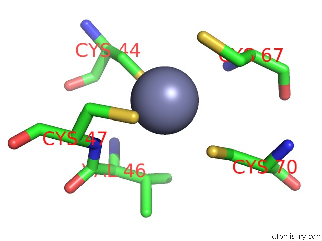
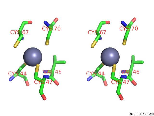
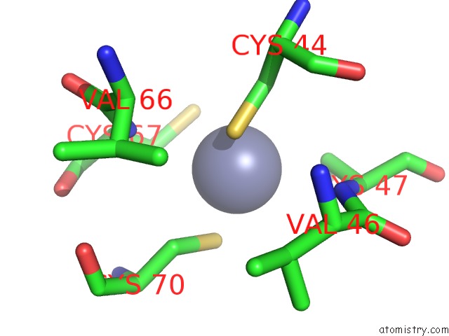
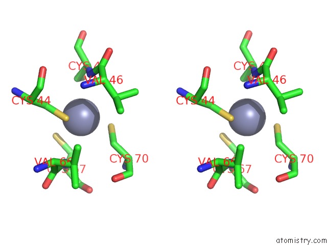
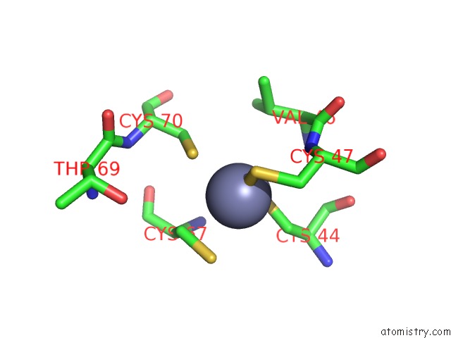
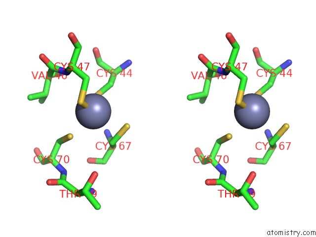
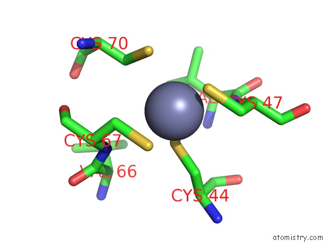
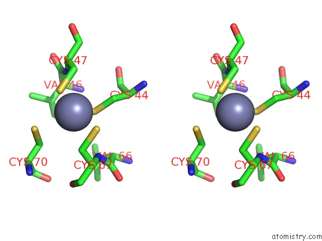
Zinc binding site 1 out of 12 in 4b86
Go back to
Zinc binding site 1 out
of 12 in the Crystal Structure of the MSL1-MSL2 Complex (3.5A)
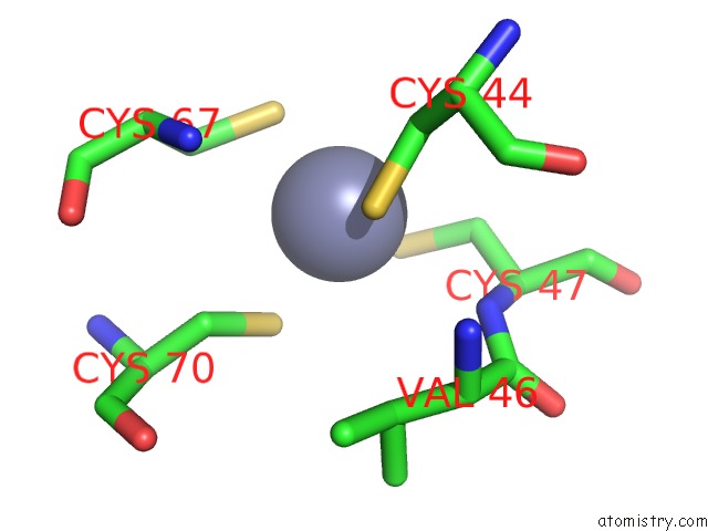
Mono view
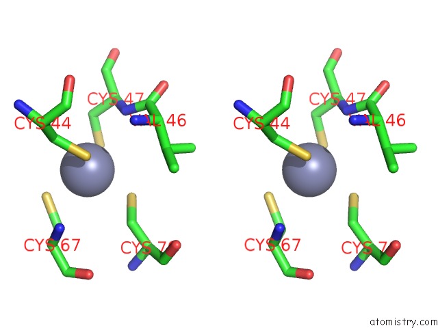
Stereo pair view
Mono view
Stereo pair view
A full contact list of Zinc with other atoms in the Zn binding
site number 1 of Crystal Structure of the MSL1-MSL2 Complex (3.5A) within 5.0Å range:
|
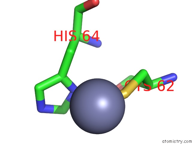
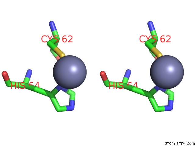
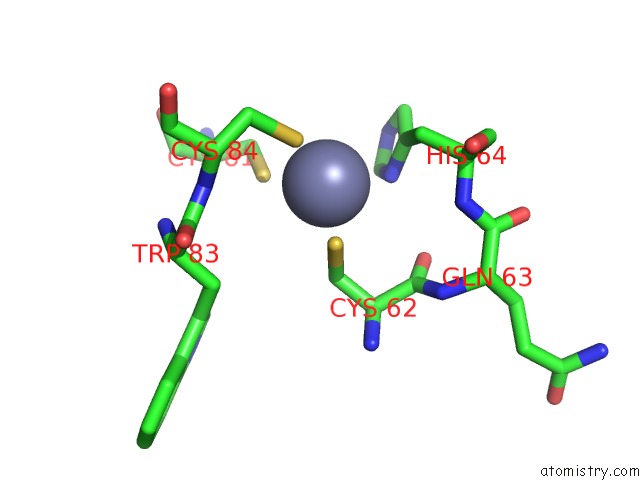
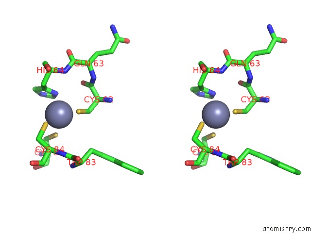
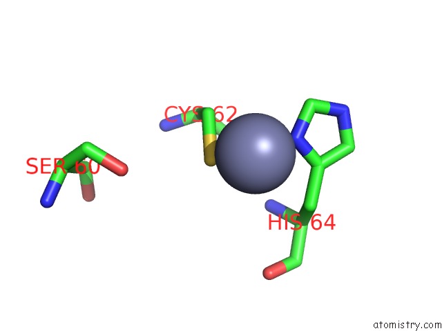
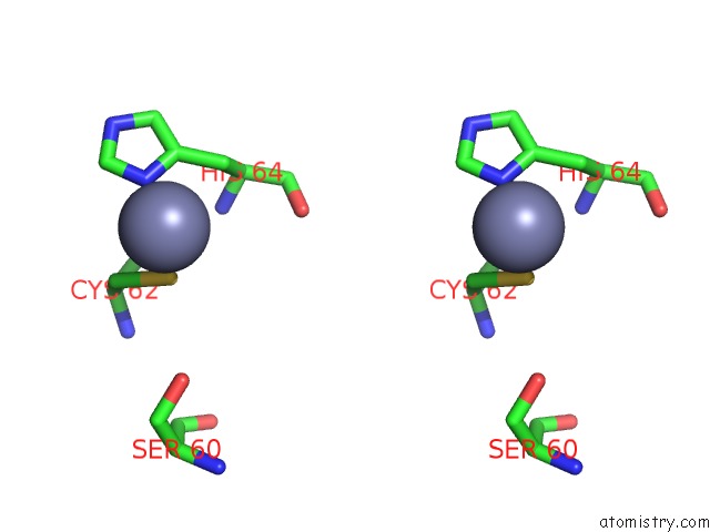
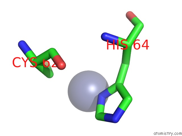
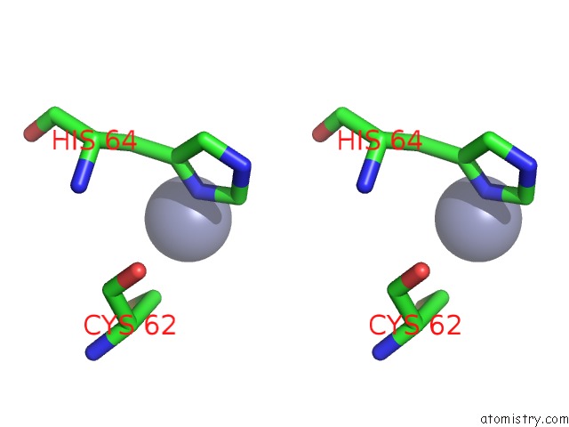
Zinc binding site 2 out of 12 in 4b86
Go back to
Zinc binding site 2 out
of 12 in the Crystal Structure of the MSL1-MSL2 Complex (3.5A)
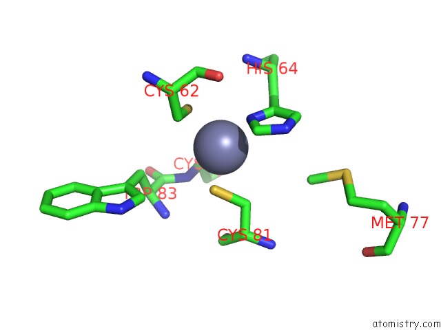
Mono view
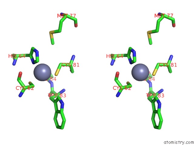
Stereo pair view
Mono view
Stereo pair view
A full contact list of Zinc with other atoms in the Zn binding
site number 2 of Crystal Structure of the MSL1-MSL2 Complex (3.5A) within 5.0Å range:
|
Zinc binding site 3 out of 12 in 4b86
Go back to
Zinc binding site 3 out
of 12 in the Crystal Structure of the MSL1-MSL2 Complex (3.5A)
Mono view
Stereo pair view
Mono view
Stereo pair view
A full contact list of Zinc with other atoms in the Zn binding
site number 3 of Crystal Structure of the MSL1-MSL2 Complex (3.5A) within 5.0Å range:
|
Zinc binding site 4 out of 12 in 4b86
Go back to
Zinc binding site 4 out
of 12 in the Crystal Structure of the MSL1-MSL2 Complex (3.5A)
Mono view
Stereo pair view
Mono view
Stereo pair view
A full contact list of Zinc with other atoms in the Zn binding
site number 4 of Crystal Structure of the MSL1-MSL2 Complex (3.5A) within 5.0Å range:
|
Zinc binding site 5 out of 12 in 4b86
Go back to
Zinc binding site 5 out
of 12 in the Crystal Structure of the MSL1-MSL2 Complex (3.5A)
Mono view
Stereo pair view
Mono view
Stereo pair view
A full contact list of Zinc with other atoms in the Zn binding
site number 5 of Crystal Structure of the MSL1-MSL2 Complex (3.5A) within 5.0Å range:
|
Zinc binding site 6 out of 12 in 4b86
Go back to
Zinc binding site 6 out
of 12 in the Crystal Structure of the MSL1-MSL2 Complex (3.5A)
Mono view
Stereo pair view
Mono view
Stereo pair view
A full contact list of Zinc with other atoms in the Zn binding
site number 6 of Crystal Structure of the MSL1-MSL2 Complex (3.5A) within 5.0Å range:
|
Zinc binding site 7 out of 12 in 4b86
Go back to
Zinc binding site 7 out
of 12 in the Crystal Structure of the MSL1-MSL2 Complex (3.5A)
Mono view
Stereo pair view
Mono view
Stereo pair view
A full contact list of Zinc with other atoms in the Zn binding
site number 7 of Crystal Structure of the MSL1-MSL2 Complex (3.5A) within 5.0Å range:
|
Zinc binding site 8 out of 12 in 4b86
Go back to
Zinc binding site 8 out
of 12 in the Crystal Structure of the MSL1-MSL2 Complex (3.5A)
Mono view
Stereo pair view
Mono view
Stereo pair view
A full contact list of Zinc with other atoms in the Zn binding
site number 8 of Crystal Structure of the MSL1-MSL2 Complex (3.5A) within 5.0Å range:
|
Zinc binding site 9 out of 12 in 4b86
Go back to
Zinc binding site 9 out
of 12 in the Crystal Structure of the MSL1-MSL2 Complex (3.5A)
Mono view
Stereo pair view
Mono view
Stereo pair view
A full contact list of Zinc with other atoms in the Zn binding
site number 9 of Crystal Structure of the MSL1-MSL2 Complex (3.5A) within 5.0Å range:
|
Zinc binding site 10 out of 12 in 4b86
Go back to
Zinc binding site 10 out
of 12 in the Crystal Structure of the MSL1-MSL2 Complex (3.5A)
Mono view
Stereo pair view
Mono view
Stereo pair view
A full contact list of Zinc with other atoms in the Zn binding
site number 10 of Crystal Structure of the MSL1-MSL2 Complex (3.5A) within 5.0Å range:
|
Reference:
E.Hallacli,
M.Lipp,
P.Georgiev,
C.Spielman,
S.Cusack,
A.Akhtar,
J.Kadlec.
MSL1-Mediated Dimerization of the Dosage Compensation Complex Is Essential For Male X-Chromosome Regulation in Drosophila. Mol.Cell V. 48 587 2012.
ISSN: ISSN 1097-2765
PubMed: 23084835
DOI: 10.1016/J.MOLCEL.2012.09.014
Page generated: Sat Oct 26 19:35:26 2024
ISSN: ISSN 1097-2765
PubMed: 23084835
DOI: 10.1016/J.MOLCEL.2012.09.014
Last articles
Mn in 5WFWMn in 5WFM
Mn in 5WF3
Mn in 5WEY
Mn in 5WEI
Mn in 5WEB
Mn in 5WEF
Mn in 5WDY
Mn in 5WE8
Mn in 5WE7