Zinc in PDB 8r1i: Human Carbonic Anhydrase II (Hcaii) in Complex with (R)-N-(3-Indol-1- Yl-2-Methyl-Propyl)-4-Sulfamoyl-Benzamide
Enzymatic activity of Human Carbonic Anhydrase II (Hcaii) in Complex with (R)-N-(3-Indol-1- Yl-2-Methyl-Propyl)-4-Sulfamoyl-Benzamide
All present enzymatic activity of Human Carbonic Anhydrase II (Hcaii) in Complex with (R)-N-(3-Indol-1- Yl-2-Methyl-Propyl)-4-Sulfamoyl-Benzamide:
4.2.1.1;
4.2.1.1;
Protein crystallography data
The structure of Human Carbonic Anhydrase II (Hcaii) in Complex with (R)-N-(3-Indol-1- Yl-2-Methyl-Propyl)-4-Sulfamoyl-Benzamide, PDB code: 8r1i
was solved by
J.Kotschy,
R.Gasper,
R.Linser,
with X-Ray Crystallography technique. A brief refinement statistics is given in the table below:
| Resolution Low / High (Å) | 32.10 / 1.46 |
| Space group | P 1 21 1 |
| Cell size a, b, c (Å), α, β, γ (°) | 42.352, 41.595, 72.243, 90, 104.34, 90 |
| R / Rfree (%) | 16.8 / 19.2 |
Zinc Binding Sites:
The binding sites of Zinc atom in the Human Carbonic Anhydrase II (Hcaii) in Complex with (R)-N-(3-Indol-1- Yl-2-Methyl-Propyl)-4-Sulfamoyl-Benzamide
(pdb code 8r1i). This binding sites where shown within
5.0 Angstroms radius around Zinc atom.
In total only one binding site of Zinc was determined in the Human Carbonic Anhydrase II (Hcaii) in Complex with (R)-N-(3-Indol-1- Yl-2-Methyl-Propyl)-4-Sulfamoyl-Benzamide, PDB code: 8r1i:
In total only one binding site of Zinc was determined in the Human Carbonic Anhydrase II (Hcaii) in Complex with (R)-N-(3-Indol-1- Yl-2-Methyl-Propyl)-4-Sulfamoyl-Benzamide, PDB code: 8r1i:
Zinc binding site 1 out of 1 in 8r1i
Go back to
Zinc binding site 1 out
of 1 in the Human Carbonic Anhydrase II (Hcaii) in Complex with (R)-N-(3-Indol-1- Yl-2-Methyl-Propyl)-4-Sulfamoyl-Benzamide
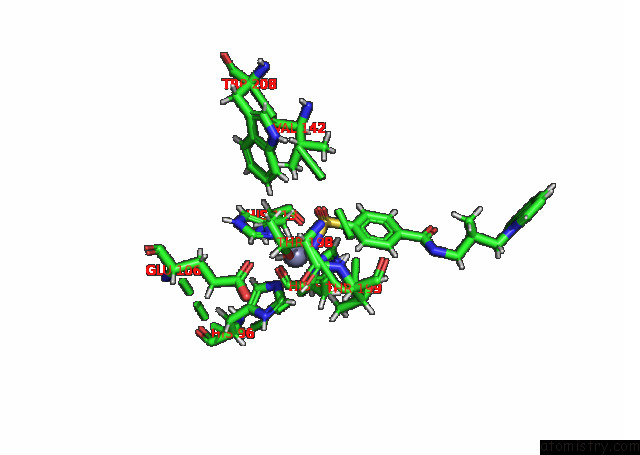
Mono view
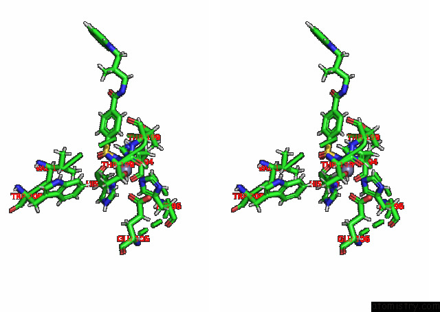
Stereo pair view
Mono view
Stereo pair view
A full contact list of Zinc with other atoms in the Zn binding
site number 1 of Human Carbonic Anhydrase II (Hcaii) in Complex with (R)-N-(3-Indol-1- Yl-2-Methyl-Propyl)-4-Sulfamoyl-Benzamide within 5.0Å range:
|
Reference:
J.Kotschy,
B.Soldner,
H.Singh,
S.K.Vasa,
R.Linser.
Microsecond Timescale Conformational Dynamics of A Small-Molecule Ligand Within the Active Site of A Protein. Angew.Chem.Int.Ed.Engl. 13947 2023.
ISSN: ESSN 1521-3773
PubMed: 37974542
DOI: 10.1002/ANIE.202313947
Page generated: Thu Oct 31 10:21:51 2024
ISSN: ESSN 1521-3773
PubMed: 37974542
DOI: 10.1002/ANIE.202313947
Last articles
Zn in 9JYWZn in 9IR4
Zn in 9IR3
Zn in 9GMX
Zn in 9GMW
Zn in 9JEJ
Zn in 9ERF
Zn in 9ERE
Zn in 9EGV
Zn in 9EGW