Zinc in PDB 7v8f: Crystal Structure of UBE2L3 Bound to Hoip RING1 Domain.
Enzymatic activity of Crystal Structure of UBE2L3 Bound to Hoip RING1 Domain.
All present enzymatic activity of Crystal Structure of UBE2L3 Bound to Hoip RING1 Domain.:
2.3.2.23; 2.3.2.31;
2.3.2.23; 2.3.2.31;
Protein crystallography data
The structure of Crystal Structure of UBE2L3 Bound to Hoip RING1 Domain., PDB code: 7v8f
was solved by
J.Liu,
Y.Wang,
L.Pan,
with X-Ray Crystallography technique. A brief refinement statistics is given in the table below:
| Resolution Low / High (Å) | 54.21 / 1.66 |
| Space group | P 1 21 1 |
| Cell size a, b, c (Å), α, β, γ (°) | 30.097, 108.412, 37.156, 90, 106.33, 90 |
| R / Rfree (%) | 17.5 / 19.8 |
Zinc Binding Sites:
The binding sites of Zinc atom in the Crystal Structure of UBE2L3 Bound to Hoip RING1 Domain.
(pdb code 7v8f). This binding sites where shown within
5.0 Angstroms radius around Zinc atom.
In total 2 binding sites of Zinc where determined in the Crystal Structure of UBE2L3 Bound to Hoip RING1 Domain., PDB code: 7v8f:
Jump to Zinc binding site number: 1; 2;
In total 2 binding sites of Zinc where determined in the Crystal Structure of UBE2L3 Bound to Hoip RING1 Domain., PDB code: 7v8f:
Jump to Zinc binding site number: 1; 2;
Zinc binding site 1 out of 2 in 7v8f
Go back to
Zinc binding site 1 out
of 2 in the Crystal Structure of UBE2L3 Bound to Hoip RING1 Domain.
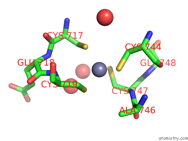
Mono view
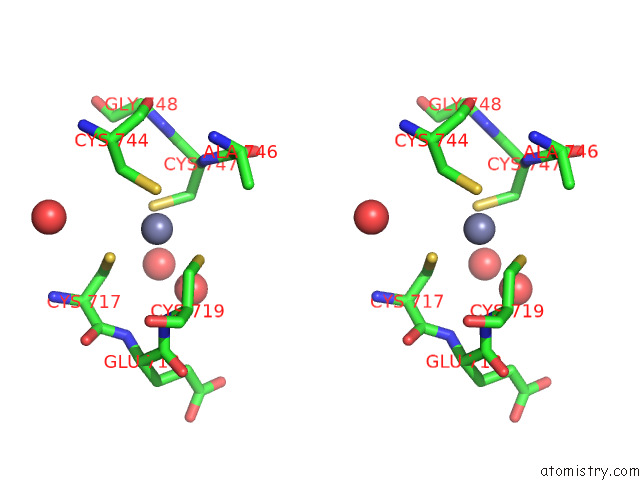
Stereo pair view
Mono view
Stereo pair view
A full contact list of Zinc with other atoms in the Zn binding
site number 1 of Crystal Structure of UBE2L3 Bound to Hoip RING1 Domain. within 5.0Å range:
|
Zinc binding site 2 out of 2 in 7v8f
Go back to
Zinc binding site 2 out
of 2 in the Crystal Structure of UBE2L3 Bound to Hoip RING1 Domain.
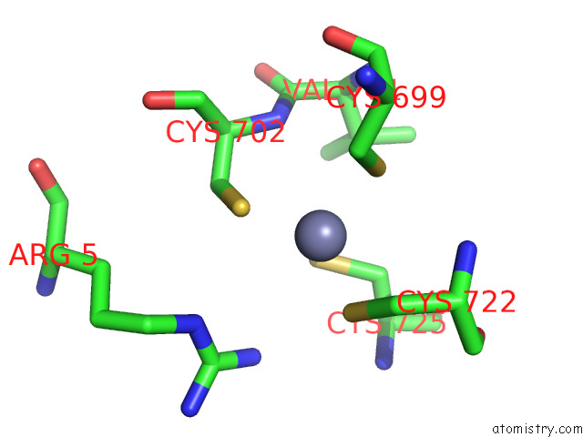
Mono view
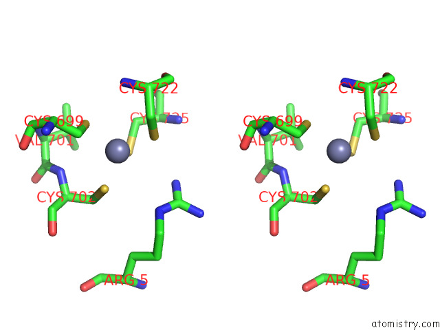
Stereo pair view
Mono view
Stereo pair view
A full contact list of Zinc with other atoms in the Zn binding
site number 2 of Crystal Structure of UBE2L3 Bound to Hoip RING1 Domain. within 5.0Å range:
|
Reference:
J.Liu,
Y.Wang,
D.Wang,
Y.Wang,
X.Xu,
Y.Zhang,
Y.Li,
M.Zhang,
X.Gong,
Y.Tang,
L.Shen,
M.Li,
L.Pan.
Mechanistic Insights Into the Subversion of the Linear Ubiquitin Chain Assembly Complex By the E3 Ligase IPAH1.4 of Shigella Flexneri. Proc.Natl.Acad.Sci.Usa V. 119 76119 2022.
ISSN: ESSN 1091-6490
PubMed: 35294289
DOI: 10.1073/PNAS.2116776119
Page generated: Wed Oct 30 12:34:59 2024
ISSN: ESSN 1091-6490
PubMed: 35294289
DOI: 10.1073/PNAS.2116776119
Last articles
Zn in 9JYWZn in 9IR4
Zn in 9IR3
Zn in 9GMX
Zn in 9GMW
Zn in 9JEJ
Zn in 9ERF
Zn in 9ERE
Zn in 9EGV
Zn in 9EGW