Zinc in PDB 7uxd: Crystal Structure of APOBEC3G Catalytic Domain Complex with Ssdna Containing 2'-Deoxy Zebularine.
Enzymatic activity of Crystal Structure of APOBEC3G Catalytic Domain Complex with Ssdna Containing 2'-Deoxy Zebularine.
All present enzymatic activity of Crystal Structure of APOBEC3G Catalytic Domain Complex with Ssdna Containing 2'-Deoxy Zebularine.:
3.5.4.38;
3.5.4.38;
Protein crystallography data
The structure of Crystal Structure of APOBEC3G Catalytic Domain Complex with Ssdna Containing 2'-Deoxy Zebularine., PDB code: 7uxd
was solved by
A.Maiti,
H.Matsuo,
with X-Ray Crystallography technique. A brief refinement statistics is given in the table below:
| Resolution Low / High (Å) | 34.51 / 1.50 |
| Space group | P 1 21 1 |
| Cell size a, b, c (Å), α, β, γ (°) | 47.489, 47.121, 52.042, 90, 103.18, 90 |
| R / Rfree (%) | 17.3 / 19.4 |
Zinc Binding Sites:
The binding sites of Zinc atom in the Crystal Structure of APOBEC3G Catalytic Domain Complex with Ssdna Containing 2'-Deoxy Zebularine.
(pdb code 7uxd). This binding sites where shown within
5.0 Angstroms radius around Zinc atom.
In total only one binding site of Zinc was determined in the Crystal Structure of APOBEC3G Catalytic Domain Complex with Ssdna Containing 2'-Deoxy Zebularine., PDB code: 7uxd:
In total only one binding site of Zinc was determined in the Crystal Structure of APOBEC3G Catalytic Domain Complex with Ssdna Containing 2'-Deoxy Zebularine., PDB code: 7uxd:
Zinc binding site 1 out of 1 in 7uxd
Go back to
Zinc binding site 1 out
of 1 in the Crystal Structure of APOBEC3G Catalytic Domain Complex with Ssdna Containing 2'-Deoxy Zebularine.
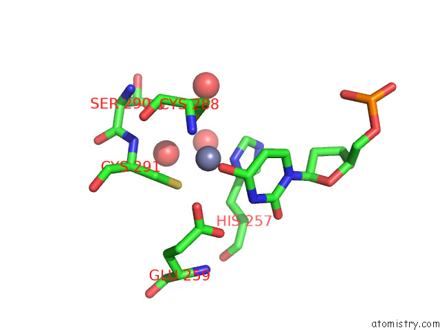
Mono view
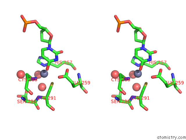
Stereo pair view
Mono view
Stereo pair view
A full contact list of Zinc with other atoms in the Zn binding
site number 1 of Crystal Structure of APOBEC3G Catalytic Domain Complex with Ssdna Containing 2'-Deoxy Zebularine. within 5.0Å range:
|
Reference:
A.Maiti,
A.K.Hedger,
W.Myint,
V.Balachandran,
J.K.Watts,
C.A.Schiffer,
H.Matsuo.
Structure of the Catalytically Active APOBEC3G Bound to A Dna Oligonucleotide Inhibitor Reveals Tetrahedral Geometry of the Transition State. Nat Commun V. 13 7117 2022.
ISSN: ESSN 2041-1723
PubMed: 36402773
DOI: 10.1038/S41467-022-34752-1
Page generated: Wed Oct 30 12:24:41 2024
ISSN: ESSN 2041-1723
PubMed: 36402773
DOI: 10.1038/S41467-022-34752-1
Last articles
Zn in 9JYWZn in 9IR4
Zn in 9IR3
Zn in 9GMX
Zn in 9GMW
Zn in 9JEJ
Zn in 9ERF
Zn in 9ERE
Zn in 9EGV
Zn in 9EGW