Zinc in PDB 7t5t: Structure of Thauera Sp. K11 Capp
Protein crystallography data
The structure of Structure of Thauera Sp. K11 Capp, PDB code: 7t5t
was solved by
R.K.Lau,
K.D.Corbett,
with X-Ray Crystallography technique. A brief refinement statistics is given in the table below:
| Resolution Low / High (Å) | 39.49 / 1.35 |
| Space group | P 42 21 2 |
| Cell size a, b, c (Å), α, β, γ (°) | 95.309, 95.309, 104.915, 90, 90, 90 |
| R / Rfree (%) | 16.2 / 16.8 |
Zinc Binding Sites:
The binding sites of Zinc atom in the Structure of Thauera Sp. K11 Capp
(pdb code 7t5t). This binding sites where shown within
5.0 Angstroms radius around Zinc atom.
In total only one binding site of Zinc was determined in the Structure of Thauera Sp. K11 Capp, PDB code: 7t5t:
In total only one binding site of Zinc was determined in the Structure of Thauera Sp. K11 Capp, PDB code: 7t5t:
Zinc binding site 1 out of 1 in 7t5t
Go back to
Zinc binding site 1 out
of 1 in the Structure of Thauera Sp. K11 Capp
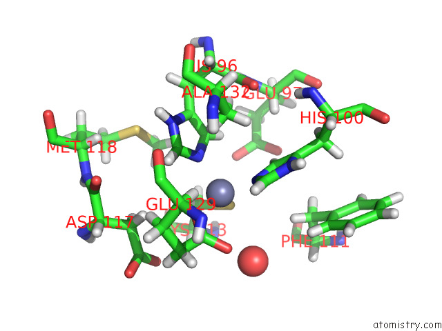
Mono view
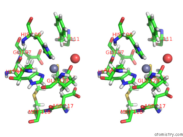
Stereo pair view
Mono view
Stereo pair view
A full contact list of Zinc with other atoms in the Zn binding
site number 1 of Structure of Thauera Sp. K11 Capp within 5.0Å range:
|
Reference:
R.K.Lau,
E.Enustun,
Y.Gu,
J.V.Nguyen,
K.D.Corbett.
A Conserved Signaling Pathway Activates Bacterial Cbass Immune Signaling in Response to Dna Damage. Embo J. V. 41 11540 2022.
ISSN: ESSN 1460-2075
PubMed: 36156805
DOI: 10.15252/EMBJ.2022111540
Page generated: Wed Oct 30 11:26:16 2024
ISSN: ESSN 1460-2075
PubMed: 36156805
DOI: 10.15252/EMBJ.2022111540
Last articles
Zn in 9JYWZn in 9IR4
Zn in 9IR3
Zn in 9GMX
Zn in 9GMW
Zn in 9JEJ
Zn in 9ERF
Zn in 9ERE
Zn in 9EGV
Zn in 9EGW