Zinc in PDB 7s68: Structure of Human PARP1 Domains (ZN1, ZN3, Wgr and Hd) Bound to A Dna Double Strand Break.
Enzymatic activity of Structure of Human PARP1 Domains (ZN1, ZN3, Wgr and Hd) Bound to A Dna Double Strand Break.
All present enzymatic activity of Structure of Human PARP1 Domains (ZN1, ZN3, Wgr and Hd) Bound to A Dna Double Strand Break.:
2.4.2.30;
Protein crystallography data
The structure of Structure of Human PARP1 Domains (ZN1, ZN3, Wgr and Hd) Bound to A Dna Double Strand Break., PDB code: 7s68
was solved by
E.Rouleau-Turcotte,
J.M.Pascal,
with X-Ray Crystallography technique. A brief refinement statistics is given in the table below:
Resolution Low / High (Å)
|
47.41 /
3.30
|
Space group
|
C 1 2 1
|
Cell size a, b, c (Å), α, β, γ (°)
|
87.208,
93.652,
119.259,
90,
106.29,
90
|
R / Rfree (%)
|
25.1 /
29.2
|
Zinc Binding Sites:
The binding sites of Zinc atom in the Structure of Human PARP1 Domains (ZN1, ZN3, Wgr and Hd) Bound to A Dna Double Strand Break.
(pdb code 7s68). This binding sites where shown within
5.0 Angstroms radius around Zinc atom.
In total 2 binding sites of Zinc where determined in the
Structure of Human PARP1 Domains (ZN1, ZN3, Wgr and Hd) Bound to A Dna Double Strand Break., PDB code: 7s68:
Jump to Zinc binding site number:
1;
2;
Zinc binding site 1 out
of 2 in 7s68
Go back to
Zinc Binding Sites List in 7s68
Zinc binding site 1 out
of 2 in the Structure of Human PARP1 Domains (ZN1, ZN3, Wgr and Hd) Bound to A Dna Double Strand Break.
Mono view
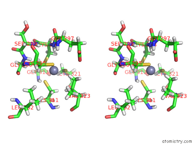
Stereo pair view
|
A full contact list of Zinc with other atoms in the Zn binding
site number 1 of Structure of Human PARP1 Domains (ZN1, ZN3, Wgr and Hd) Bound to A Dna Double Strand Break. within 5.0Å range:
probe
|
atom
|
residue
|
distance (Å)
|
B
|
Occ
|
A:Zn401
b:221.0
occ:1.00
|
SG
|
A:CYS298
|
2.2
|
103.5
|
1.0
|
SG
|
A:CYS295
|
2.3
|
178.6
|
1.0
|
SG
|
A:CYS311
|
2.3
|
130.5
|
1.0
|
SG
|
A:CYS321
|
2.3
|
104.3
|
1.0
|
HB2
|
A:CYS295
|
2.8
|
157.2
|
1.0
|
HG21
|
A:VAL323
|
2.9
|
148.7
|
1.0
|
CB
|
A:CYS295
|
2.9
|
130.6
|
1.0
|
H
|
A:CYS298
|
3.1
|
181.8
|
1.0
|
HB3
|
A:CYS295
|
3.1
|
157.2
|
1.0
|
HB3
|
A:CYS311
|
3.2
|
176.3
|
1.0
|
HB3
|
A:CYS298
|
3.2
|
160.4
|
1.0
|
CB
|
A:CYS311
|
3.3
|
146.5
|
1.0
|
HB2
|
A:CYS311
|
3.4
|
176.3
|
1.0
|
HB3
|
A:CYS321
|
3.4
|
185.9
|
1.0
|
CB
|
A:CYS298
|
3.4
|
133.2
|
1.0
|
CB
|
A:CYS321
|
3.4
|
154.5
|
1.0
|
HB
|
A:VAL323
|
3.6
|
137.4
|
1.0
|
CG2
|
A:VAL323
|
3.7
|
123.5
|
1.0
|
HB2
|
A:CYS321
|
3.7
|
185.9
|
1.0
|
HG23
|
A:VAL323
|
3.8
|
148.7
|
1.0
|
N
|
A:CYS298
|
3.9
|
151.1
|
1.0
|
H
|
A:GLY300
|
4.0
|
163.1
|
1.0
|
HB2
|
A:GLU297
|
4.1
|
203.8
|
1.0
|
HB2
|
A:CYS298
|
4.1
|
160.4
|
1.0
|
CB
|
A:VAL323
|
4.2
|
114.1
|
1.0
|
CA
|
A:CYS298
|
4.2
|
135.3
|
1.0
|
H
|
A:GLY313
|
4.3
|
160.2
|
1.0
|
H
|
A:GLU297
|
4.4
|
169.5
|
1.0
|
CA
|
A:CYS295
|
4.4
|
135.3
|
1.0
|
HD11
|
A:LEU302
|
4.4
|
162.1
|
1.0
|
HG22
|
A:VAL323
|
4.4
|
148.7
|
1.0
|
H
|
A:SER299
|
4.5
|
147.5
|
1.0
|
HA3
|
A:GLY313
|
4.6
|
161.7
|
1.0
|
HG11
|
A:VAL323
|
4.6
|
152.6
|
1.0
|
HA
|
A:CYS295
|
4.6
|
162.9
|
1.0
|
HD12
|
A:LEU302
|
4.6
|
162.1
|
1.0
|
CA
|
A:CYS311
|
4.7
|
122.5
|
1.0
|
N
|
A:GLY300
|
4.8
|
135.5
|
1.0
|
HA3
|
A:GLY300
|
4.8
|
157.6
|
1.0
|
CA
|
A:CYS321
|
4.8
|
151.7
|
1.0
|
N
|
A:SER299
|
4.8
|
122.5
|
1.0
|
C
|
A:CYS298
|
4.8
|
151.8
|
1.0
|
OE1
|
A:GLU297
|
5.0
|
134.1
|
1.0
|
CG1
|
A:VAL323
|
5.0
|
126.7
|
1.0
|
CB
|
A:GLU297
|
5.0
|
169.4
|
1.0
|
H
|
A:VAL323
|
5.0
|
152.6
|
1.0
|
HA
|
A:CYS311
|
5.0
|
147.5
|
1.0
|
HA
|
A:CYS298
|
5.0
|
162.8
|
1.0
|
|
Zinc binding site 2 out
of 2 in 7s68
Go back to
Zinc Binding Sites List in 7s68
Zinc binding site 2 out
of 2 in the Structure of Human PARP1 Domains (ZN1, ZN3, Wgr and Hd) Bound to A Dna Double Strand Break.
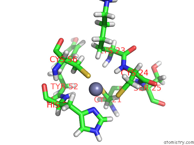
Mono view
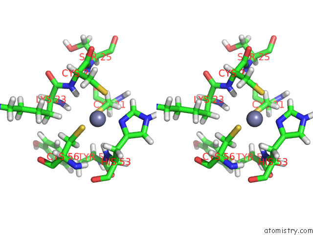
Stereo pair view
|
A full contact list of Zinc with other atoms in the Zn binding
site number 2 of Structure of Human PARP1 Domains (ZN1, ZN3, Wgr and Hd) Bound to A Dna Double Strand Break. within 5.0Å range:
probe
|
atom
|
residue
|
distance (Å)
|
B
|
Occ
|
A:Zn402
b:155.4
occ:1.00
|
ND1
|
A:HIS53
|
2.0
|
106.7
|
1.0
|
SG
|
A:CYS24
|
2.3
|
138.4
|
1.0
|
SG
|
A:CYS56
|
2.3
|
140.8
|
1.0
|
SG
|
A:CYS21
|
2.4
|
117.7
|
1.0
|
HB2
|
A:HIS53
|
2.4
|
183.8
|
1.0
|
HB2
|
A:CYS56
|
2.8
|
175.1
|
1.0
|
CG
|
A:HIS53
|
2.9
|
140.4
|
1.0
|
H
|
A:HIS53
|
2.9
|
162.2
|
1.0
|
H
|
A:CYS24
|
3.0
|
165.2
|
1.0
|
CB
|
A:HIS53
|
3.1
|
152.7
|
1.0
|
CB
|
A:CYS56
|
3.1
|
145.5
|
1.0
|
CE1
|
A:HIS53
|
3.1
|
125.8
|
1.0
|
HB3
|
A:CYS24
|
3.3
|
174.6
|
1.0
|
HB3
|
A:CYS56
|
3.4
|
175.1
|
1.0
|
HE1
|
A:HIS53
|
3.4
|
151.5
|
1.0
|
CB
|
A:CYS24
|
3.4
|
145.1
|
1.0
|
HB2
|
A:CYS21
|
3.5
|
165.2
|
1.0
|
HB3
|
A:LYS23
|
3.5
|
185.9
|
1.0
|
CB
|
A:CYS21
|
3.5
|
137.3
|
1.0
|
N
|
A:HIS53
|
3.6
|
134.7
|
1.0
|
HB3
|
A:CYS21
|
3.7
|
165.2
|
1.0
|
N
|
A:CYS24
|
3.8
|
137.2
|
1.0
|
HB3
|
A:HIS53
|
3.8
|
183.8
|
1.0
|
CA
|
A:HIS53
|
4.0
|
136.9
|
1.0
|
CD2
|
A:HIS53
|
4.0
|
146.4
|
1.0
|
NE2
|
A:HIS53
|
4.1
|
147.0
|
1.0
|
HB2
|
A:CYS24
|
4.2
|
174.6
|
1.0
|
CA
|
A:CYS24
|
4.2
|
117.9
|
1.0
|
H
|
A:LYS23
|
4.3
|
146.1
|
1.0
|
H
|
A:CYS56
|
4.3
|
155.4
|
1.0
|
CB
|
A:LYS23
|
4.4
|
154.5
|
1.0
|
HA
|
A:TYR52
|
4.5
|
115.8
|
1.0
|
CA
|
A:CYS56
|
4.5
|
158.4
|
1.0
|
HA
|
A:HIS53
|
4.6
|
164.8
|
1.0
|
N
|
A:CYS56
|
4.8
|
129.1
|
1.0
|
C
|
A:TYR52
|
4.8
|
98.9
|
1.0
|
C
|
A:LYS23
|
4.8
|
148.8
|
1.0
|
H
|
A:SER25
|
4.8
|
168.4
|
1.0
|
HD2
|
A:HIS53
|
4.8
|
176.2
|
1.0
|
HG3
|
A:LYS23
|
4.9
|
206.0
|
1.0
|
HE2
|
A:HIS53
|
4.9
|
176.9
|
1.0
|
HG2
|
A:LYS23
|
4.9
|
206.0
|
1.0
|
CA
|
A:CYS21
|
4.9
|
116.5
|
1.0
|
HA
|
A:CYS24
|
4.9
|
142.0
|
1.0
|
N
|
A:LYS23
|
5.0
|
121.3
|
1.0
|
HA
|
A:CYS56
|
5.0
|
190.5
|
1.0
|
C
|
A:HIS53
|
5.0
|
108.3
|
1.0
|
CA
|
A:LYS23
|
5.0
|
116.7
|
1.0
|
HB3
|
A:TYR52
|
5.0
|
155.8
|
1.0
|
|
Reference:
E.Rouleau-Turcotte,
D.B.Krastev,
S.J.Pettitt,
C.J.Lord,
J.M.Pascal.
Captured Snapshots of PARP1 in the Active State Reveal the Mechanics of PARP1 Allostery. Mol.Cell V. 82 2939 2022.
ISSN: ISSN 1097-2765
PubMed: 35793673
DOI: 10.1016/J.MOLCEL.2022.06.011
Page generated: Wed Oct 30 10:37:54 2024
|